| Journal of Hematology, ISSN 1927-1212 print, 1927-1220 online, Open Access |
| Article copyright, the authors; Journal compilation copyright, J Hematol and Elmer Press Inc |
| Journal website https://jh.elmerpub.com |
Case Report
Volume 15, Number 5, October 2026, pages 255-259

Measles Pneumonia After Allogeneic Hematopoietic Stem Cell Transplantation
Marco Alejandro Jimenez-Ochoaa, c, Maria Margarita Contreras-Serratosa, Elizabeth Urbina-Escalantea, Maria Gabriela Pardo-Escobedoa, Erendira Pacheco-Zavalaa, Victor Adrian Cortes-Moralesb
aMexican Social Security Institute, National Medical Center Siglo XXI, Bone Marrow Transplantation Unit, Mexico City, Mexico
bMexican Social Security Institute, National Medical Center Siglo XXI, Medical Research Unit in Immunochemistry, Mexico City, Mexico
cCorresponding Author: Marco Alejandro Jimenez-Ochoa, Mexican Social Security Institute, National Medical Center Siglo XXI, Bone Marrow Transplantation Unit, 06720 Mexico City, Mexico
Manuscript submitted August 4, 2026, accepted September 2, 2026, published online October 31, 2026
Short title: Measles After Allogeneic Transplantation
doi: https://doi.org/10.14740/jh2234
| Abstract | ▴Top |
Measles is a highly contagious viral infection that is rarely reported after allogeneic hematopoietic stem cell transplantation (HSCT), with only a limited number of cases described in the literature. We report the case of a 46-year-old woman who developed laboratory-confirmed measles complicated by atypical pneumonia 3 years after human leukocyte antigen (HLA)-identical sibling allogeneic HSCT for acute lymphoblastic leukemia. Her post-transplant course had been complicated by steroid-refractory chronic graft-versus-host disease (GVHD), successfully treated with ruxolitinib, which had been discontinued only a few months before presentation. She was admitted with fever, severe odynophagia, and diffuse oral mucosal erythema, leading to an initial diagnosis of chronic GVHD flare and treatment with corticosteroids and empirical antimicrobials. During hospitalization, she rapidly developed hypoxemic respiratory failure and a characteristic cephalocaudal maculopapular rash. Reverse transcription polymerase chain reaction (RT-PCR) of a throat swab confirmed measles virus infection. Corticosteroids were discontinued, and the patient received supportive care and intravenous immunoglobulin (IVIG), with complete clinical and radiologic recovery. Notably, despite adequate quantitative immune reconstitution, including preserved CD4+ and CD8+ T-cell counts and normal immunoglobulin levels, she remained susceptible to measles because she was not yet eligible for measles-mumps-rubella (MMR) vaccination according to current post-transplant recommendations. This case highlights the diagnostic challenge of distinguishing measles from chronic GVHD in transplant recipients, emphasizes that quantitative immune recovery does not necessarily reflect pathogen-specific immunity, and underscores the importance of maintaining a high index of suspicion and performing early molecular testing during community outbreaks.
Keywords: Measles; Graft-versus-host disease; Pneumonia; Hematopoietic stem cell transplantation
| Introduction | ▴Top |
Measles is a highly contagious viral disease caused by a single-stranded, negative-sense RNA virus belonging to the Paramyxoviridae family and the Morbillivirus genus. Transmission occurs primarily through airborne spread via aerosols and respiratory droplets, with a secondary attack rate of up to 90% among susceptible contacts [1].
Following an incubation period of approximately 10–14 days, the disease typically presents with high fever, cough, coryza, conjunctivitis, and a cephalocaudal maculopapular rash. Although most cases are self-limiting, severe complications and death may occur, particularly in immunocompromised individuals [2].
In Mexico, the current measles outbreak began in February 2025, with a sustained increase in incidence from five cases per 100,000 population in 2025 to eight cases per 100,000 population in 2026. This resurgence has been attributed to multiple factors, including declining vaccination coverage, heterogeneity in population immunity, and several sociodemographic factors that have facilitated the re-introduction and transmission of the virus [3, 4].
Measles outbreaks disproportionately affect vulnerable populations, including pregnant women, immunocompromised patients, and malnourished children, in whom the risk of severe disease and complications is substantially increased [5]. In patients with hematologic malignancies, transplant candidates and other immunosuppressive conditions, higher rates of pneumonia, encephalitis, and fatal outcomes have been reported [6].
The largest published series of measles in hematopoietic stem cell transplant recipients corresponds to the outbreak reported in Sao Paulo, Brazil, in which eight allogeneic hematopoietic stem cell transplant recipients developed laboratory-confirmed measles during a community outbreak; only one patient developed pneumonia [7].
Despite the global resurgence of measles, reports in hematopoietic stem cell transplant recipients remain exceedingly rare. Consequently, the clinical spectrum, diagnostic approach, and optimal management of these profoundly immunosuppressed patients remain poorly defined. We present the case of a recipient of an allogeneic hematopoietic stem cell transplantation (HSCT) who developed measles during the outbreak in Mexico. The disease manifested shortly after the discontinuation of prolonged immunosuppressive therapy, with an initial presentation that closely mimicked a flare of graft-versus-host disease (GVHD).
| Case Report | ▴Top |
Investigations
A 46-year-old female nursing assistant with a history of CD20-positive common B-cell acute lymphoblastic leukemia, considered high-risk because of her age at diagnosis, achieved complete remission following hyperfractionated cyclophosphamide, vincristine, doxorubicin, and dexamethasone (hyper-CVAD) chemotherapy and subsequently underwent a human leukocyte antigen (HLA)-identical sibling allogeneic HSCT. She received a myeloablative conditioning regimen consisting of busulfan, cyclophosphamide, and fludarabine (BuCyFlu), GVHD prophylaxis with cyclosporine and methotrexate. The early post-transplant course was complicated by grade II acute gastrointestinal GVHD during immunosuppression tapering, which responded completely to a short course of corticosteroids. She also developed pure red cell aplasia secondary to major ABO incompatibility, which resolved spontaneously around day +100. Subsequently, she developed steroid-refractory chronic mucocutaneous GVHD and was treated with ruxolitinib for 1 year, achieving complete disease control and permanent discontinuation of immunosuppressive therapy 3 years after transplantation. At the time of the case presentation, the patient had isolated oral lichen planus, classified as mild chronic GVHD, managed with symptomatic treatment only. Because of persistent chronic GVHD and the recent withdrawal of immunosuppressive therapy, she did not receive the measles-mumps-rubella (MMR) vaccine as part of the post-transplant revaccination schedule. Both the patient and the donor had no history of measles infection prior to transplantation. Three years and 3 months after transplantation, she presented to the emergency department with a 3-day history of fever up to 39 °C, predominantly occurring at night, accompanied by chills, severe odynophagia with inability to tolerate oral intake, and occasional diarrhea.
During the first 24 h of hospitalization, her clinical condition deteriorated with the development of cough, acute hypoxemic respiratory failure with a peripheral oxygen saturation of 75%, and the appearance of an intensely pruritic erythematous maculopapular rash. The rash initially involved the face and trunk and subsequently progressed in a cephalocaudal distribution to the upper and lower extremities over the following 3 days (Fig. 1). No Koplik spots were observed.
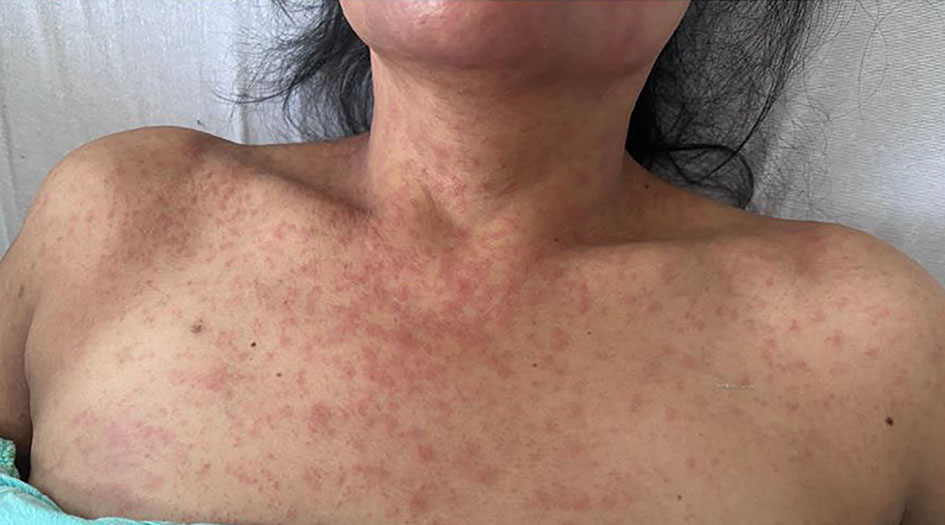 Click for large image | Figure 1. Diffuse erythematous maculopapular rash involving the anterior neck and upper chest. |
Diagnosis
Complete blood count and serum chemistry revealed no significant abnormalities. Polymerase chain reaction (PCR) assays for cytomegalovirus, Epstein–Barr virus, adenovirus, and parvovirus B19 were all negative. Lymphocyte subset analysis demonstrated adequate T-cell immune reconstitution, with CD3+, CD4+, and CD8+ counts of 2,495, 990, and 1,206 cells/µL, respectively (CD4+/CD8+ ratio, 0.82) and preserved IgG levels of 2,040 mg/dL (reference value: 700–1,600 mg/dL), IgM of 238 mg/dL (reference value: 40–240 mg/dL), and IgA of 68 mg/dL (reference value: 65–400 mg/dL).
High-resolution computed tomography of the chest demonstrated ground-glass opacities in the right lower lobe, consistent with atypical pneumonia (Fig. 2).
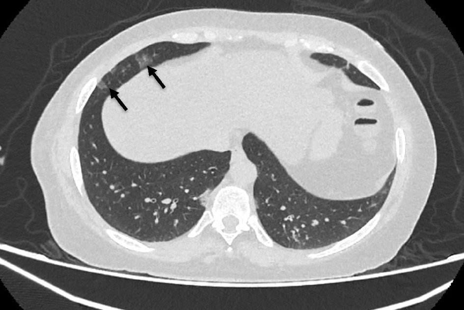 Click for large image | Figure 2. High-resolution chest computed tomography showing patchy ground-glass opacities involving the right lower lobe, findings consistent with viral pneumonia. The arrows indicate the sites of pulmonary involvement. |
The rapid clinical deterioration, respiratory involvement, and characteristic rash made a GVHD flare an unlikely primary cause of her presentation. Although the patient initially denied any epidemiologic history of contact with individuals with measles, the ongoing nationwide outbreak and the high clinical suspicion prompted testing for measles and rubella by reverse transcription polymerase chain reaction (RT-PCR) performed on a throat swab specimen. The assay was positive for measles virus and negative for rubella, with results available within 24 h.
Based on the clinical, radiologic, and molecular findings, a diagnosis of measles virus infection complicated by pneumonia, pharyngitis, and exanthematous dermatitis was established.
Treatment
Given the suspicion of an upper respiratory tract infection associated with a possible flare of oral GVHD, empirical antimicrobial therapy and intravenous methylprednisolone (1 mg/kg/day) were initiated. Following confirmation of measles, methylprednisolone was discontinued after two doses, as a GVHD flare was ruled out. Supportive care included supplemental oxygen, initially delivered via nasal cannula and subsequently escalated to a mask for 3 days because of increasing oxygen requirements. Given her history of allogeneic HSCT, pulmonary involvement, and severe clinical presentation, intravenous immunoglobulin (IVIG, 0.5 g/kg) was administered. Additional supportive treatment included inhaled β2-agonists, inhaled corticosteroids, antihistamines, and symptomatic care.
Follow-up and outcomes
The patient’s clinical course was favorable. From the sixth day of hospitalization, the rash gradually resolved and oxygen requirements progressively decreased. She was discharged on hospital day 8, afebrile, without the need for supplemental oxygen, and with near-complete resolution of the cutaneous lesions.
At the 2-week follow-up visit, chest computed tomography demonstrated complete resolution of the pulmonary abnormalities. A directed epidemiologic interview conducted after the diagnosis revealed that one of her children had been in close contact with a confirmed case of measles, an exposure that had not been initially reported by the patient. Three months after the infection, the patient remains completely asymptomatic, without the need for immunosuppressive therapy, recurrence of GVHD, or respiratory or cutaneous sequelae.
| Discussion | ▴Top |
Reports of measles after allogeneic HSCT remain exceedingly rare. To our knowledge, only one case series and a few isolated case reports have been published. The largest published series included eight allogeneic HSCT recipients during a community outbreak in Brazil, with pneumonia occurring in only one patient. A summary of the published cases is presented in Table 1. Across the published reports, measles in allogeneic HSCT recipients has consistently presented with fever and a generalized maculopapular rash, frequently accompanied by respiratory manifestations ranging from upper respiratory tract symptoms to interstitial pneumonia. Most patients were diagnosed during community or nosocomial outbreaks. IVIG was administered in the majority of patients with moderate or severe disease, whereas pneumonia was uncommon and generally associated with greater immunosuppression. Overall, clinical outcomes were favorable, with only one reported death occurring in a patient who developed rapidly progressive measles pneumonia during active immunosuppressive therapy [7–9].
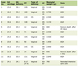 Click to view | Table 1. Compilation of Cases With Measles After Allogeneic HSCT |
Our case expands the currently available literature in several clinically relevant aspects. In contrast to previously reported patients, measles developed more than 3 years after allogeneic HSCT, at a time when the patient had recently discontinued prolonged immunosuppressive therapy for steroid-refractory chronic mucocutaneous GVHD. She presented with fever, severe odynophagia, inability to tolerate oral intake, and diffuse oral mucosal erythema, making an exacerbation of chronic GVHD the most likely initial diagnosis. This diagnostic impression was further supported by previous reports demonstrating that viral infections, particularly cytomegalovirus, herpes simplex virus, parvovirus B19, and more recently severe acute respiratory syndrome coronavirus 2 (SARS-CoV-2), may trigger or exacerbate GVHD [10]. Consequently, empirical corticosteroids and antimicrobial therapy were initiated while an appropriate microbiologic workup was performed. However, the subsequent development of a characteristic cephalocaudal maculopapular rash and atypical pneumonia, together with the ongoing national measles outbreak, prompted RT-PCR testing despite the patient’s initial denial of a known epidemiologic exposure, ultimately establishing the correct diagnosis.
In the cohort reported by Machado et al [7], severe measles was associated with patients receiving high-dose immunosuppressive therapy, a finding consistent with the fatal case described by Lee et al, who developed rapidly progressive measles pneumonia while under active immunosuppression [8]. In contrast, our patient developed severe measles more than 3 years after allogeneic HSCT, at a time when she had achieved apparently adequate quantitative immune reconstitution. Two months before the infection, peripheral blood lymphocyte subsets demonstrated normal CD4+ and CD8+ T-cell recovery, and serum immunoglobulin levels were preserved, with an elevated IgG level likely reflecting chronic immune activation related to previous GVHD. Nevertheless, these parameters do not necessarily indicate protective immunity against measles. Garcia-Garrido et al demonstrated that measles-specific humoral immunity progressively declines after allogeneic HSCT, with seroprotection decreasing from 91% before transplantation to 86% at 3 months and only 61% at 1 year after transplantation. This decline was particularly pronounced among recipients of myeloablative conditioning regimens, suggesting that adequate quantitative immune reconstitution does not necessarily translate into preserved pathogen-specific immune memory [11].
Current guidelines from the American Society for Transplantation and Cellular Therapy, the Infectious Diseases Society of America, and the National Comprehensive Cancer Network recommend administration of the live-attenuated MMR vaccine only in allogeneic HSCT recipients who are at least 24 months post-transplantation, have no active chronic GVHD, are no longer receiving systemic immunosuppressive therapy, and have not received IVIG within the preceding 8–11 months. Similarly, Carpenter et al proposed the practical “2-1-8 rule”: vaccination should be considered only after 2 years post-transplantation, 1 year off systemic immunosuppression, and 8 months after the last IVIG administration [12, 13]. Our patient did not meet any of these eligibility criteria, as ruxolitinib had been discontinued only a few months before presentation and she was still undergoing clinical evaluation for a possible recurrence of chronic GVHD.
Consequently, deferral of MMR vaccination was consistent with current recommendations rather than a missed opportunity for prevention. Although the available evidence suggests that the MMR vaccine is generally safe in carefully selected HSCT recipients, data remain limited for patients with recent immunosuppression or active GVHD. Moreover, rare cases of vaccine-associated measles have been reported in transplant recipients, although these have generally followed a mild and self-limited clinical course.
Measles can cause two main patterns of pulmonary involvement: interstitial pneumonia and the more severe giant cell pneumonia, or Hecht pneumonia, which is particularly associated with immunocompromised patients and may lead to respiratory failure and death.
Histologically, Hecht pneumonia is characterized by extensive alveolar epithelial injury and multinucleated giant cells with viral inclusions; however, these findings may overlap with interstitial pneumonia and may require immunohistochemistry or electron microscopy for confirmation [14]. In our patient, bronchoscopy and lung biopsy were not performed because of her favorable clinical improvement.
Learning points
This case highlights several important clinical lessons. First, measles should be considered in the differential diagnosis of allogeneic HSCT recipients presenting with fever, mucosal involvement, and rash during community outbreaks, regardless of the time elapsed since transplantation. Second, recent withdrawal of immunosuppressive therapy and a history of chronic GVHD may lead to an initial misdiagnosis, delaying recognition of measles. Third, adequate quantitative immune reconstitution does not necessarily reflect preserved pathogen-specific immunity, emphasizing the need for timely molecular testing and strict adherence to current revaccination recommendations in this vulnerable population. Finally, during measles outbreaks, particular emphasis should be placed on preventive measures to protect HSCT recipients from exposure, including the use of face masks and avoidance of contact with suspected or confirmed cases, given their increased risk of severe disease.
Acknowledgments
We would like to acknowledge the epidemiology department of our hospital for their collaboration, and all the residents of hematology of our center who were involved in the treatment of this patient.
Financial Disclosure
None to declare.
Conflict of Interest
None to declare.
Informed Consent
Written informed consent was obtained from the patient for the publication of this case report and any accompanying images.
Author Contributions
Jimenez-Ochoa and Contreras-Serratos conceived the presented idea. Urbina-Escalante and Jimenez-Ochoa developed the theory and performed the analysis. Pardo-Escobedo, Pacheco-Zavala, and Cortes-Morales supervised the findings of this work and checked the English translation. All authors discussed the results and contributed to the final manuscript.
Data Availability
The authors declare that data supporting the findings of this study are available within the article.
| References | ▴Top |
- Moss WJ. Measles. Lancet. 2017;390(10111):2490-2502.
doi pubmed - Mathis AD, Raines K, Filardo TD, Wiley N, Leung J, Rota PA, Martinez D, et al. Measles update - United States, January 1-April 17, 2025. MMWR Morb Mortal Wkly Rep. 2025;74(14):232-238.
doi pubmed - Sandoval-Pinto E, Cremades R, Perez-Gomez HR, Rivera-Avila RC, Barbosa CAD, Premkumar L, Sierra-Diaz E, et al. Spatiotemporal dynamics of the 2025-2026 measles outbreak in Mexico: Jalisco as the emerging epicenter and the impact of intensive immunization efforts. Pathogens. 2026;15(6).
doi pubmed - De Arcos-Jimenez JC, Martinez-Ayala P, Fernandez-Diaz OF, Sanchez-Enriquez S, Vargas-Becerra PN, Lopez-Yanez AM, Damian-Negrete R, et al. Social determinants and outbreak dynamics of the 2025 measles epidemic in Mexico: a nationwide analysis of linked surveillance data. Viruses. 2026;18(2).
doi pubmed - Do LAH, Mulholland K. Measles 2025. N Engl J Med. 2025;393(24):2447-2458.
doi pubmed - Kreiger-Benson E, Gelb B, Neumann HJ, Hochman S, Lighter J, Mehta SA. Measles outbreak risk assessment for transplant candidates and recipients. Am J Transplant. 2021;21(1):338-343.
doi pubmed - Machado CM, Goncalves FB, Pannuti CS, Dulley FL, de Souza VA. Measles in bone marrow transplant recipients during an outbreak in Sao Paulo, Brazil. Blood. 2002;99(1):83-87.
doi pubmed - Lee DG, Yoo JH, Choi JH, Choi SM, Park SH, Kim YJ, Kim DW, et al. A fatal case of measles pneumonia complicating an adult recipient of hemopoietic stem cell transplantation during the nationwide epidemic in Korea. Int J Infect Dis. 2006;10(5):410-411.
doi pubmed - Ge YL, Zhai XW, Zhu YF, Wang XS, Xia AM, Li YF, Zeng M. Measles outbreak in pediatric hematology and oncology patients in Shanghai, 2015. Chin Med J (Engl). 2017;130(11):1320-1326.
doi pubmed - Florenzo B, Martin S, Saavedra A. Reactivated chronic graft-versus-host disease following SARS-CoV-2 infection. JAAD Case Rep. 2022;26:76-78.
doi pubmed - Garcia Garrido HM, van Aalst M, Schinkel J, Koen G, Defoer JM, Hazenberg MD, Nur E, et al. Early loss of immunity against measles following allogeneic hematopoietic stem cell transplantation. Am J Hematol. 2019;94(10):E270-E272.
doi pubmed - Chang SY, Bisht A, Faysman K, Schiller GJ, Uslan DZ, Multani A. Vaccine-associated measles in a hematopoietic cell transplant recipient: case report and comprehensive review of the literature. Open Forum Infect Dis. 2021;8(8):ofab326.
doi pubmed - Carpenter PA, Englund JA. How I vaccinate blood and marrow transplant recipients. Blood. 2016;127(23):2824-2832.
doi pubmed - Moussallem TM, Guedes F, Fernandes ER, Pagliari C, Lancellotti CL, de Andrade HF, Jr., Duarte MI. Lung involvement in childhood measles: severe immune dysfunction revealed by quantitative immunohistochemistry. Hum Pathol. 2007;38(8):1239-1247.
doi pubmed
This article is distributed under the terms of the Creative Commons Attribution 4.0 International License (CC BY 4.0), which permits unrestricted use, distribution, and reproduction in any medium, including commercial use, provided the original work is properly cited.
Journal of Hematology is published by Elmer Press Inc.