| Journal of Hematology, ISSN 1927-1212 print, 1927-1220 online, Open Access |
| Article copyright, the authors; Journal compilation copyright, J Hematol and Elmer Press Inc |
| Journal website https://jh.elmerpub.com |
Case Report
Volume 15, Number 5, October 2026, pages 248-254

A Case of Classic Hodgkin Lymphoma With Biventricular Myocardial Involvement
Justin T. Blackmana, b, i, Jessica Blackmanc, Markus Bernardus Sikkela, d, e, f, g, Tara Baetzc, h
aIsland Medical Program, Faculty of Medicine, University of British Columbia, Vancouver, BC, Canada
bUniversity of Victoria, PO Box 1700 STN CSC, Victoria, BC V8W 2Y2, Canada
cMalignant Hematology, British Columbia Cancer Agency, Victoria, BC, Canada
dDivision of Cardiology, Royal Jubilee Hospital, Victoria, BC, Canada
eDivision of Cardiology, University of British Columbia, Vancouver, BC, Canada
fCenter for Cardiovascular Innovation, University of British Columbia, Vancouver, BC, Canada
gDivision of Medical Sciences, University of Victoria, Victoria, BC, Canada
hDivision of Medicine, University of British Columbia, Vancouver, BC, Canada
iCorresponding Author: Justin T. Blackman, University of Victoria, PO Box 1700 STN CSC, Victoria, BC V8W 2Y2, Canada
Manuscript submitted June 23, 2026, accepted September 2, 2026, published online October 31, 2026
Short title: CHL With Myocardial Involvement
doi: https://doi.org/10.14740/jh2226
| Abstract | ▴Top |
Pericardial disease occurs in approximately 5–25% of patients with classic Hodgkin lymphoma, representing a minority overall but occurring more frequently in patients with bulky mediastinal disease, whereas direct myocardial invasion is rarely reported. We report the case of a 31-year-old male with International Prognostic Score (IPS)-7 score of 4, stage IVB classic Hodgkin lymphoma, nodular sclerosis subtype, who initially presented with multiple mediastinal masses with tumor infiltration of both ventricles—the first documented case of biventricular myocardial involvement with multiple cardiac masses. Myocardial involvement was diagnosed clinically and radiographically on concordant multimodality imaging and supported by response to lymphoma-directed therapy; no myocardial biopsy was performed, and histologic confirmation of classic Hodgkin lymphoma was obtained from a left axillary lymph node excisional biopsy. He received one cycle of cyclophosphamide, doxorubicin, vincristine, and prednisone (CHOP) chemotherapy prior to pathology confirmation, then six cycles of brentuximab plus doxorubicin, vinblastine, and dacarbazine (A + AVD). A marked morphologic response followed the single initial CHOP cycle, and a significant metabolic response was seen after two subsequent A + AVD cycles. He completed therapy without cardiac rupture despite major tumor burden, though during treatment he required hospitalization for a syncopal episode associated with extensive great vessel thrombosis and paradoxical hypotensive bradycardia. The thrombosis was considered most consistent with an advanced-stage, early-treatment lymphoma-associated hypercoagulable state, although a contribution from antecedent vascular compression could not be excluded.
Keywords: Cardio-oncology; Hodgkin disease; Lymphoma; Antineoplastic combined chemotherapy protocols
| Introduction | ▴Top |
Direct myocardial involvement in classic Hodgkin lymphoma (cHL) is exceptionally uncommon. cHL is itself an uncommon malignancy with an incidence rate of approximately 0.98 per 100,000 individuals globally [1]. Though pericardial disease occurs in 5–25% of cHL cases [2], few cases of direct myocardial invasion have ever been documented [2–7]. We report the case of a 31-year-old man diagnosed with International Prognostic Score (IPS)-7 score of 4, stage IVB, cHL, nodular sclerosis subtype, who initially presented with multiple mediastinal masses and tumor infiltration of the myocardium in both ventricles; to our knowledge, this is the first-ever report of cHL with biventricular myocardial involvement and multiple cardiac masses. Myocardial involvement was diagnosed clinically and radiographically on concordant multimodality imaging and was supported by response to lymphoma-directed therapy; no myocardial biopsy was performed, and histologic confirmation of cHL was obtained from a left axillary lymph node excisional biopsy.
| Case Report | ▴Top |
A previously healthy 31-year-old male patient presented with a 6-month history of drenching night sweats, fevers, palpable left-sided axillary adenopathy, and severe chest pain worse when supine. On day 0, he was admitted following computed tomography (CT) findings of several large mediastinal masses with the largest measuring 10.3 cm in maximum dimension (Fig. 1a). He presented with leukocytosis and relative lymphocytopenia (neutrophils: 11.09 × 10⁹/L (reference range: 2.00–6.00 × 10⁹/L), eosinophils: 0.92 × 10⁹/L (reference range: < 0.45 × 10⁹/L), basophils: 0.88 × 10⁹/L (reference range: < 0.10 × 10⁹/L), lymphocytes: 0.96 × 10⁹/L (reference range: 1.00–4.00 × 10⁹/L)), elevated C-reactive protein of 18.1 mg/L (reference range: < 3.0 mg/L), and undetectable troponin I (< 3 ng/L; reference range: < 53 ng/L).
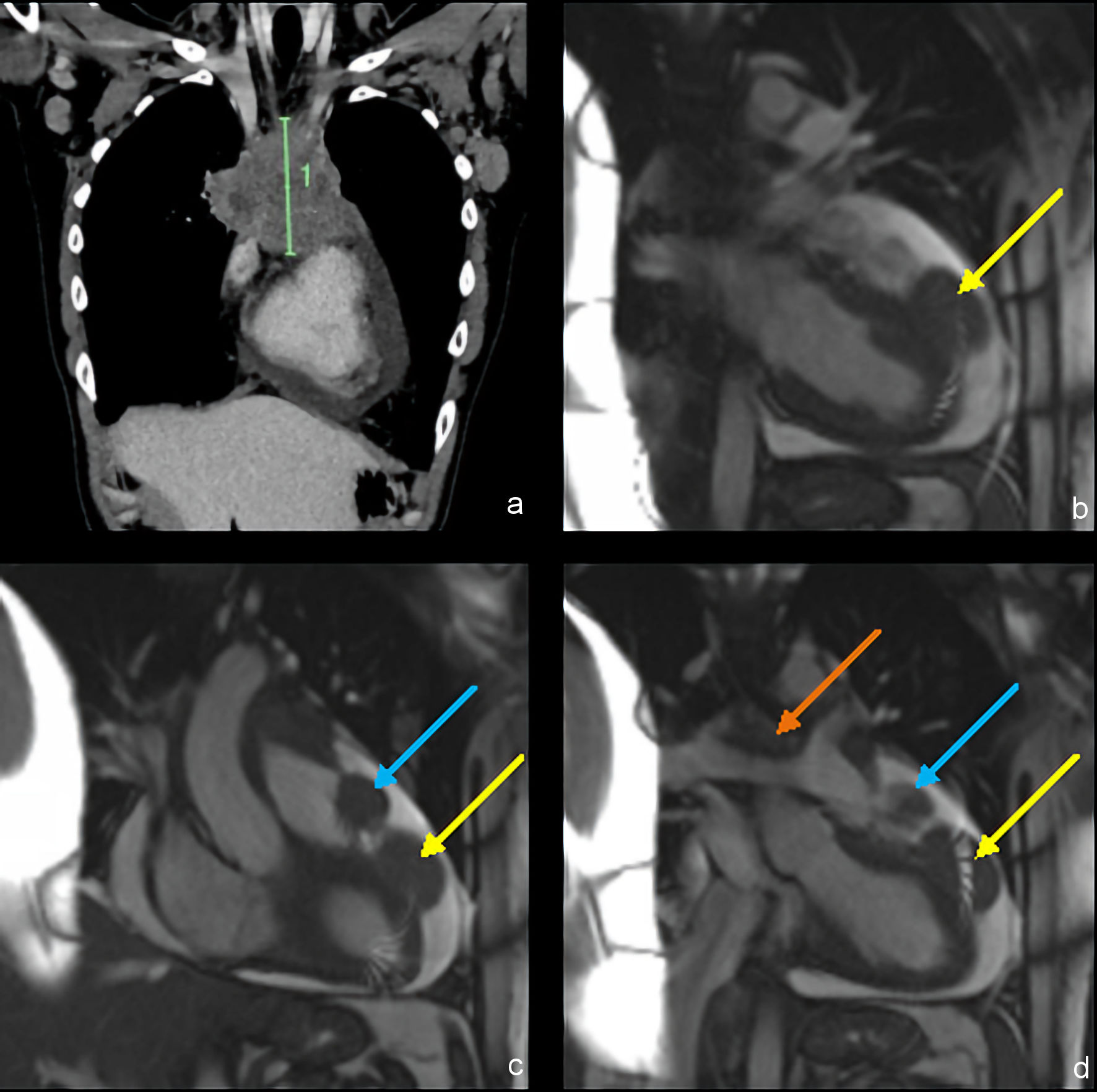 Click for large image | Figure 1. Contrast-enhanced CT (a) and contrast-enhanced ECG-gated MRI (b–d) imaging of mediastinal masses before therapy. (a) Coronal image shows a large anterior mediastinal mass (10.3 cm in length). (b) Coronal image shows a left ventricular mass with middle and superficial myocardial involvement (yellow arrow). (c) Coronal image shows a right ventricular outflow tract mass (blue arrow). (d) Coronal image shows a great vessel mass compressing the pulmonary arteries (orange arrow). CT: computed tomography; ECG: electrocardiogram; MRI: magnetic resonance imaging. |
Transthoracic echocardiogram on day 1 reported a left ventricular ejection fraction of 45% with global hypokinesis, no valvular disease, and no evidence of tamponade. There was a large circumferential pericardial effusion measuring up to 1.8 cm. He underwent cardiac magnetic resonance imaging (MRI) on day 2, which found three distinct cardiac masses, among others on the chest wall. One mass was contiguous with the epicardial surface of the mid-to-apical anterior wall of the left ventricle and measured 2.8 × 4.6 × 2.7 cm in size (Fig. 1b); this mass involved the epicardial and mid-myocardial layers and extended into the pericardial space but did not involve the parietal pericardium. A second similar mass arising along the left lateral aspect of the right ventricular outflow tract measuring 2.7 × 2.4 × 2.6 cm was also noted (Fig. 1c). The second mass similarly did not invade the parietal pericardium but did involve the wall of the outflow tract. A third mass was noted in the anterior mediastinum with ill-defined margins but clearly encasing the ascending aorta and compressing the main pulmonary artery, the right pulmonary artery, and the superior vena cava (Fig. 1d). Several nodules along the epicardial surface of the right ventricular outflow tract were also identified.
He underwent a left axillary lymph node core biopsy and bone marrow biopsy on day 2. The bone marrow biopsy was negative for lymphoma involvement. Preliminary lymph node pathology became available on day 10 and demonstrated an aggressive lymphoma with features suggestive of cHL; however, the morphology was not definitive and raised the possibility of mediastinal gray zone lymphoma. A left axillary lymph node excisional biopsy was therefore performed on day 12. Given his ongoing symptoms and substantial tumor burden, including several large intracardiac masses, systemic therapy was initiated before final pathology was available to avoid further treatment delay. Cyclophosphamide, doxorubicin, vincristine, and prednisone (CHOP) chemotherapy was selected as an empiric regimen for its activity across several aggressive lymphoma subtypes while the diagnosis remained uncertain, with the plan to modify therapy as needed once definitive pathology became available.
One cycle of CHOP was initiated on day 14. One week of bisoprolol and telemetry monitoring were also initiated at this time due to the risks of dysrhythmia and/or cardiac rupture [8], during which he remained hemodynamically stable with no dysrhythmias or signs of hemodynamic compromise.
Fluorodeoxyglucose (FDG) positron emission tomography (PET) CT imaging was performed just prior to chemotherapy on day 14 (Fig. 2a), which noted maximum standardized uptake value (SUV) of 10.8 for the left ventricular mass, and 10.4 for the right ventricular mass; the overall Deauville score was 5. Repeat echocardiogram on day 21 noted the left ventricular tumor had decreased by 1.5 cm in diameter. This and all subsequent echocardiograms showed restored left ventricular function with ejection fraction ≥ 55%.
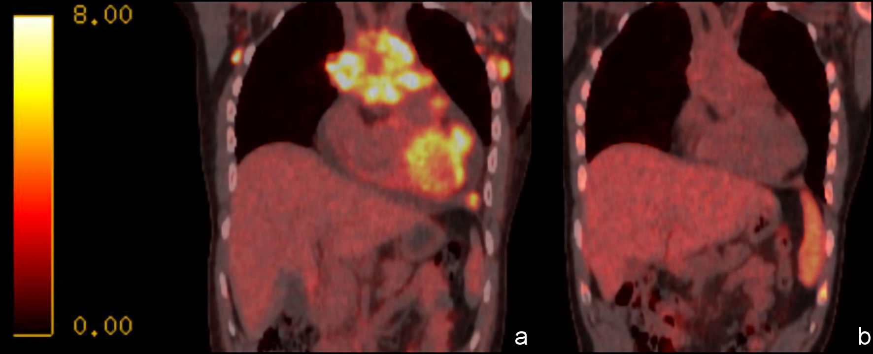 Click for large image | Figure 2. FDG PET/CT coronal images of the mediastinal masses, with SUV scale. Coronal images show (a) significant lesion metabolic activity prior to chemotherapy, and (b) significant reduction in activity following one cycle of CHOP and two cycles of A + AVD. A + AVD: brentuximab plus doxorubicin, vinblastine, and dacarbazine; CHOP: cyclophosphamide, doxorubicin, vincristine, and prednisone; CT: computed tomography; FDG: fluorodeoxyglucose; PET: positron emission tomography; SUV: standardized uptake value. |
Final pathology from the left axillary excisional lymph node biopsy became available on day 24 and confirmed cHL, nodular sclerosis subtype. He was subsequently staged as IVB based on the multiple myocardial masses and the presence of B symptoms, with a Hasenclever international prognostic score (IPS)-7 of 4. In light of the definitive pathologic findings, the treatment plan was revised to six full cycles of brentuximab plus doxorubicin, vinblastine, and dacarbazine (A + AVD) therapy [9], without counting the preceding empiric CHOP cycle toward the planned course of definitive therapy. He was discharged from hospital on day 25 with outpatient medical oncology follow-up arranged.
Cycle one day 1 A + AVD was initiated on day 36. On day 40 he experienced a syncopal episode and presented to the emergency department complaining of non-pleuritic, non-positional, left-sided chest pain at rest. He was afebrile and hypotensive but bradycardic with a heart rate of 48 beats per minute (bpm). Arterial blood gas indicated low oxyhemoglobin of 47.7% (reference range: 90.00–95.0%), physiologic pH, and mildly elevated lactate of 1.6 mmol/L (reference range: < 1.2 mmol/L). A CT pulmonary angiogram on the same day found a small thrombus of the right pulmonary artery and extensive free-floating thrombus within the superior vena cava measuring 5 cm long. This CT also demonstrated marked interval improvement in the cardiac masses and complete resolution of the pericardial effusion since treatment initiation, with the left ventricular mass reduced to 3.3 cm in maximum dimension (from 4.6 cm upon initial imaging), and the right ventricular outflow tract mass reduced to 2.0 cm (from 2.7 cm). There were no electrocardiogram (ECG) changes beyond sinus bradycardia. Troponin I was undetectable (< 3 ng/L; reference range: < 53 ng/L). No contributory metabolic derangements or other laboratory investigations were detected. He was admitted and treated with systemic anticoagulation therapy and intravenous fluids. His condition improved promptly with heart rate increasing to 85 bpm. He maintained normal sinus rhythm while on telemetry throughout the remainder of this hospitalization, and was discharged on day 56. Repeat FDG PET/CT imaging on day 91, immediately prior to cycle three of A + AVD (Fig. 2b) demonstrated significant metabolic and morphologic response to therapy, with a new SUV max of 2.3 across the cardiac-involved tumors. The original 10.3 cm anterior mediastinal mass had nearly completely resolved with residual mass measuring 1.1 cm in largest dimension. The previously noted large pericardial effusion had completely resolved. A tiny focus of residual activity in the anterior mediastinal mass suggested residual Deauville 4 activity.
He went on to complete the remainder of the six planned cycles of curative intent A + AVD on an outpatient basis without further cardiovascular complication. His last chemotherapy infusion (cycle six day 15) occurred on day 251. Repeat FDG PET/CT imaging on day 308, subsequent to completion of all treatment cycles, noted a Deauville score of 2, grossly normal heart chamber sizes, normal caliber great vessels, myocardial metabolic activity comparable to that of the background blood pool, and no pericardial effusion.
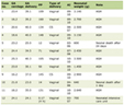
The chronology of the patient’s diagnostic evaluation, treatment, and follow-up is summarized in Table 1.
 Click to view | Table 1. Timeline of Diagnostic Evaluation, Treatment, and Follow-Up |
| Discussion | ▴Top |
A search of PubMed and Google Scholar for publications from January 2022 through May 2026 was conducted on June 21, 2026, using the leading search terms “Hodgkin” and “case,” and each of three modifiers: “cardiac,” “heart,” and “myocardium.” This search built on the methodology and findings of Kligfeld et al [2], who identified seven reported cases of myocardial involvement by Hodgkin lymphoma in the literature as of August 2023, and reported an eighth case. Our updated search identified two additional cases: one describing interventricular septal involvement [7], and another describing interatrial septum involvement with sinus node dysfunction [10].
To our knowledge, this is the 11th report of myocardial involvement of cHL in the English literature [2–7, 10, 11], the second report of cHL with ventricular myocardial involvement [7], and the first-ever report of cHL with biventricular myocardial involvement with multiple cardiac masses. Table 2 [2, 3, 5–7, 10, 11] summarizes the cardiac structures involved, presenting features, diagnostic approaches, treatments, and outcomes in selected previously reported cases.
Click to view | Table 2. Summary of Selected Published Cases of Cardiac Involvement by Classic Hodgkin Lymphoma |
Among confirmed cases of cHL with direct myocardial involvement, diagnosis has generally relied on concordant multimodality imaging together with histologic confirmation from an extracardiac site, rather than direct myocardial biopsy, consistent with the approach used in the present case. When cardiac-region tissue sampling has been attempted, it has proved technically challenging. In one reported case, an initial transcatheter biopsy of an intracardiac mass was non-diagnostic, with confirmation ultimately obtained from a pericardial-region specimen rather than transmural myocardium [11]. Treatment in all confirmed cases consisted of systemic chemotherapy without surgical resection of the cardiac lesion; however, one patient additionally required pericardiocentesis for a symptomatic effusion [2]. Outcomes were generally favorable, with resolution or marked improvement of cardiac findings following treatment, although one older report from 1989 described recurrence at 18 months [3].
The cardiac structures involved in previously reported cases have predominantly included the right atrium, interatrial septum, and adjacent great veins [2, 5, 10, 11], with interventricular septal involvement described in one case [7]. In contrast, our patient had involvement of the left ventricular free wall and right ventricular outflow tract, with no atrial or interatrial septal involvement. To our knowledge, this anatomic pattern has not previously been reported and, together with the biventricular distribution of disease, distinguishes the present case from prior reports.
Cardiac arrhythmias have been reported in association with myocardial or atrial involvement by cHL, including sinus node dysfunction from disease adjacent to the sinoatrial node [10] and supraventricular tachycardia attributed to mass effect involving the pulmonary veins [5]. In our patient, the masses did not involve the atria, interatrial septum, or cardiac conduction system, and the episode of bradycardia occurred without atrioventricular block, making direct nodal infiltration less likely. The episode instead coincided with acute superior vena caval and pulmonary arterial thrombosis and hypoxemia, although the precise mechanism underlying the bradycardia cannot be determined from the available data.
Thrombosis has only rarely been reported in association with myocardial involvement by cHL. One prior case described intra-atrial neoplastic thrombosis, representing tumor-infiltrated thrombus rather than bland clot, with recurrence at 18 months [3]. Cardiac rupture due to myocardial infiltration by lymphoma has also been associated with local hemorrhage and thrombus formation at the site of tissue destruction [8]. In contrast, our patient developed an extensive free-floating thrombus involving the superior vena cava and right pulmonary artery during a period of marked regression of the cardiac and mediastinal masses on treatment, without evidence of tumor infiltration into the thrombus or cardiac rupture. We therefore considered this more consistent with the hypercoagulable state associated with advanced-stage lymphoma early in treatment [12] rather than a direct consequence of tumor infiltration or mass effect, though a contribution from vascular compression by the antecedent mediastinal mass cannot be excluded. To our knowledge, this thrombotic presentation has not previously been described in this setting.
Conclusions
We report the successful treatment of a case of stage IVB cHL, nodular sclerosis subtype, which initially presented with biventricular myocardial involvement. Although myocardial tissue was not biopsied, biventricular myocardial involvement was supported by concordant multimodality imaging and its marked response to lymphoma-directed therapy, with cHL confirmed histologically from a left axillary lymph node excisional biopsy. Despite significant tumor burden within the myocardium, standard dose cycles of CHOP and subsequently A + AVD were tolerated without cardiac rupture. The myocardial masses were responsive to treatment, with morphologic and functional improvement within 1 week of therapy and marked morphologic and metabolic improvement within 3 months. In this patient, the myocardial masses and a large pericardial effusion ultimately resolved with systemic chemotherapy alone, without indication for urgent pericardial drainage or surgical resection. This case illustrates the utility of multimodality imaging for diagnosing and following myocardial involvement by cHL and demonstrates that even extensive biventricular involvement may respond rapidly to lymphoma-directed systemic therapy. It also highlights that cardiovascular complications arising during treatment may have mechanisms other than progressive direct cardiac tumor involvement or structural cardiac complications.
Acknowledgments
None to declare.
Financial Disclosure
No funding was received for this research.
Conflict of Interest
None to declare.
Informed Consent
Written informed consent was obtained from the patient for publication of this case report and any accompanying images.
Author Contributions
Justin T. Blackman: Writing – original draft, editing, data curation, and visualization. Jessica Blackman: Conceptualization, writing – original draft, editing, and data curation. Markus B. Sikkel: Writing – review and editing, supervision. Tara Baetz: Conceptualization, writing – review and editing, project administration. All authors read, edited, and agreed to the published version of the manuscript.
Ethics Statement
The research reported in this paper adhered to the Tri-Council Policy Statement: Ethical Conduct for Research Involving Humans (TCPS 2) guidelines and was exempted from a formal Research Ethics Board review.
Data Availability
The data that support the findings of this study are available on request from the corresponding author. The data are not publicly available due to privacy or ethical restrictions.
| References | ▴Top |
- Huang J, Pang WS, Lok V, Zhang L, Lucero-Prisno DE, 3rd, Xu W, Zheng ZJ, et al. Incidence, mortality, risk factors, and trends for Hodgkin lymphoma: a global data analysis. J Hematol Oncol. 2022;15(1):57.
doi pubmed - Kligfeld H, Wynn SM, Hermanson K, Ma TM, Suh E, Hemenway CS. An atypical presentation of Hodgkin lymphoma invading the myocardium. J Pediatr Hematol Oncol. 2023;45(6):349-351.
doi pubmed - Domergue J, Donadio D, Ferriere M, Ouaknine R, Simony J, Pujol H. Inaugural intra-atrial neoplastic thrombosis in stage II Hodgkin's disease. Eur J Surg Oncol. 1989;15(3):279-281.
pubmed - Munck JN, Bosq J, Girinsky T, da Costa L, Carde P. Primary involvement of the heart in Hodgkin's disease: prolonged survival in a patient. Eur J Med. 1993;2(8):508-509.
pubmed - Korecki J, Kaminski KA, Lisowska A, Musial WJ. Supraventricular tachycardia and pulmonary hypertension at the presentation of Hodgkin's disease. Acta Cardiol. 2005;60(6):655-657.
doi pubmed - Amirimoghaddam Z, Khoddami M, Nayeri ND, Molaee S. Hodgkin's lymphoma presenting with heart failure: a case report. J Med Case Rep. 2010;4:14.
doi pubmed - Priessnitz J, Biringen E, Khoury F, Bergin C. Treatment of myocardial infiltration of hodgkin’s lymphoma: a catch-22. JACC. 2025;85(12_Supplement):3907.
doi - Molajo AO, McWilliam L, Ward C, Rahman A. Cardiac lymphoma: an unusual case of myocardial perforation—clinical, echocardiographic, haemodynamic and pathological features. Eur Heart J. 1987;8(5):549-552.
doi pubmed - Ansell SM, Radford J, Connors JM, Dlugosz-Danecka M, Kim WS, Gallamini A, Ramchandren R, et al. Overall survival with brentuximab vedotin in stage III or IV Hodgkin's lymphoma. N Engl J Med. 2022;387(4):310-320.
doi pubmed - Marascu GE, Schmidt M, Vidlescu R, Manciu M, Cinteza E, Vatasescu RG. Sinus node dysfunction in a young patient with Hodgkin lymphoma: a case report. Eur Heart J Case Rep. 2025;9(8):ytaf360.
doi pubmed - Zoma JM, Aggarwal S, Gabali A, Kobayashi D. A rare case of primary cardiac Hodgkin lymphoma in a child. Cardiol Young. 2020;30(6):866-868.
doi pubmed - Borchmann S, Muller H, Hude I, Fuchs M, Borchmann P, Engert A. Thrombosis as a treatment complication in Hodgkin lymphoma patients: a comprehensive analysis of three prospective randomized German Hodgkin Study Group (GHSG) trials. Ann Oncol. 2019;30(8):1329-1334.
doi pubmed
This article is distributed under the terms of the Creative Commons Attribution 4.0 International License (CC BY 4.0), which permits unrestricted use, distribution, and reproduction in any medium, including commercial use, provided the original work is properly cited.
Journal of Hematology is published by Elmer Press Inc.