Journal of Hematology, ISSN 1927-1212 print, 1927-1220 online, Open Access
Article copyright, the authors; Journal compilation copyright, J Hematol and Elmer Press Inc
Journal website https://jh.elmerpub.com

Case Report

Volume 15, Number 5, October 2026, pages 248-254


A Case of Classic Hodgkin Lymphoma With Biventricular Myocardial Involvement

Figures

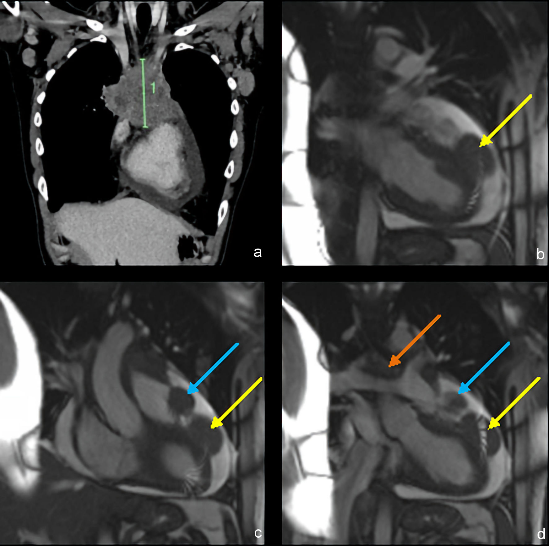
↓  Figure 1. Contrast-enhanced CT (a) and contrast-enhanced ECG-gated MRI (b–d) imaging of mediastinal masses before therapy. (a) Coronal image shows a large anterior mediastinal mass (10.3 cm in length). (b) Coronal image shows a left ventricular mass with middle and superficial myocardial involvement (yellow arrow). (c) Coronal image shows a right ventricular outflow tract mass (blue arrow). (d) Coronal image shows a great vessel mass compressing the pulmonary arteries (orange arrow). CT: computed tomography; ECG: electrocardiogram; MRI: magnetic resonance imaging.
Figure 1.
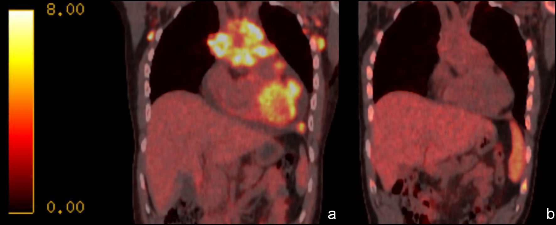
↓  Figure 2. FDG PET/CT coronal images of the mediastinal masses, with SUV scale. Coronal images show (a) significant lesion metabolic activity prior to chemotherapy, and (b) significant reduction in activity following one cycle of CHOP and two cycles of A + AVD. A + AVD: brentuximab plus doxorubicin, vinblastine, and dacarbazine; CHOP: cyclophosphamide, doxorubicin, vincristine, and prednisone; CT: computed tomography; FDG: fluorodeoxyglucose; PET: positron emission tomography; SUV: standardized uptake value.
Figure 2.

Tables

↓  Table 1. Timeline of Diagnostic Evaluation, Treatment, and Follow-Up
 
DayClinical event
A + AVD: brentuximab plus doxorubicin, vinblastine, and dacarbazine; CHOP: cyclophosphamide, doxorubicin, vincristine, and prednisone; CT: computed tomography; FDG: fluorodeoxyglucose; LVEF: left ventricular ejection fraction; MRI: magnetic resonance imaging; PET: positron emission tomography.
Day 0Initial presentation, CT, and hospital admission
Day 1Initial transthoracic echocardiogram: ejection fraction of 45%, pericardial effusion up to 1.8 cm
Day 2Cardiac MRI
Left axillary lymph node core biopsy performed
Bone marrow biopsy performed; negative for lymphoma involvement
Day 10Preliminary biopsy pathology became available: aggressive B-cell lymphoma with features suggestive of classic Hodgkin lymphoma; morphology was not definitive and raised the possibility of mediastinal gray zone lymphoma. Bone marrow negative for lymphoma involvement
Day 12Left axillary lymph node excisional biopsy
Day 14Initial FDG PET/CT
Empiric CHOP, cycle 1 day 1
Day 21Repeat echocardiogram: LVEF 55% and 1.5-cm reduction in left ventricular tumor diameter
Day 24Final excisional biopsy pathology confirmed classic Hodgkin lymphoma, nodular sclerosis subtype; definitive staging and IPS-7 assessment completed.
Day 25Discharged from initial hospitalization with outpatient medical oncology follow-up arranged
Day 36A + AVD, cycle 1 day 1
Day 40Syncopal episode prompting emergency presentation; right pulmonary artery thrombus and extensive superior vena cava thrombosis identified. Admitted to hospital.
Repeat CT: marked reduction in cardiac masses, complete resolution of pericardial effusion
Day 55Discharged from second hospitalization
Day 91Repeat FDG PET/CT
Day 251Final chemotherapy infusion, A + AVD cycle 6 day 15
Day 308Post-treatment FDG PET/CT


↓  Table 2. Summary of Selected Published Cases of Cardiac Involvement by Classic Hodgkin Lymphoma
 
Source (year)Age/sexConfirmation of diagnosisCardiac involvementPresenting featureTreatmentOutcome
F: female; FDG: fluorodeoxyglucose; LV: left ventricle; LVEF: left ventricular ejection fraction; M: male; PET: positron emission tomography; RVOT: right ventricular outflow tract; SVC: superior vena cava; PA: pulmonary artery; SVT: supraventricular tachycardia.
[3] (1989)——Intra-atrialNeoplastic intra-atrial thrombosis—Recurrence at 18 months
[5] (2005)27/FMediastinoscopy and lymph node biopsyLeft atrium; pulmonary vein compressionSVT, heart failure, pulmonary hypertensionChemotherapy not otherwise specifiedCardiac findings and arrhythmia improved
[6] (2010)8/FCervical lymph node biopsyDiffuse cardiac tissue and septumHeart failure, pericardial effusionDoxorubicin/bleomycin/vincristine/dacarbazineNo evidence of disease at 1 year
[11] (2020)14/FDiagnostic pericardial-region core biopsy after nondiagnostic transcatheter biopsyRight atrial mass extending into SVC and left innominate veinDyspnea; tricuspid inflow obstructionDoxorubicin/bleomycin/vincristine/etoposide/cyclophosphamide/prednisoneMass decreased; PET became FDG-negative
[2] (2023)18/MMediastinal mass biopsyBilateral atrial walls and right atrial chamberChest pain, large pericardial effusion, tamponade physiologyDoxorubicin/bleomycin/vinblastine/dacarbazine + pericardiocentesis + radiotherapyComplete remission > 3 years
[7] (2025)34/MLymph node biopsyInterventricular septum; global LV dysfunctionSinus tachycardia, LVEF 36%Doxorubicin/vinblastine/dacarbazine/brentuximabLVEF improved from 36% to 47%
[10] (2025)17/MCervical lymph node biopsySuperior right atrial wall and interatrial septumSinus node dysfunctionDoxorubicin/vincristine/etoposide/methylprednisolone, followed by vincristine/dacarbazine/cyclophosphamide/methylprednisoloneArrhythmia resolution by day 21; dimensional and metabolic regression within 2 months
Presented case31/MAxillary lymph node excisional biopsyLV free wall and RVOTChest pain, reduced LVEF, pericardial effusion; later syncope/bradycardia with SVC/PA thrombosisDoxorubicin/vincristine/cyclophosphamide/prednisone, followed by doxorubicin/vinblastine/dacarbazine/brentuximabNo evidence of disease at 1 year