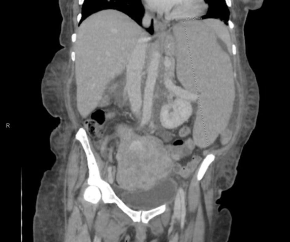
↓ Figure 1. Coronal view of CT
chest/abdomen/pelvis showing significant splenomegaly, measuring 23.5 cm in craniocaudal dimension.
Enlarged, lobulated uterus with partially calcified and heterogenous fibroids are also seen. CT:
computed tomography.
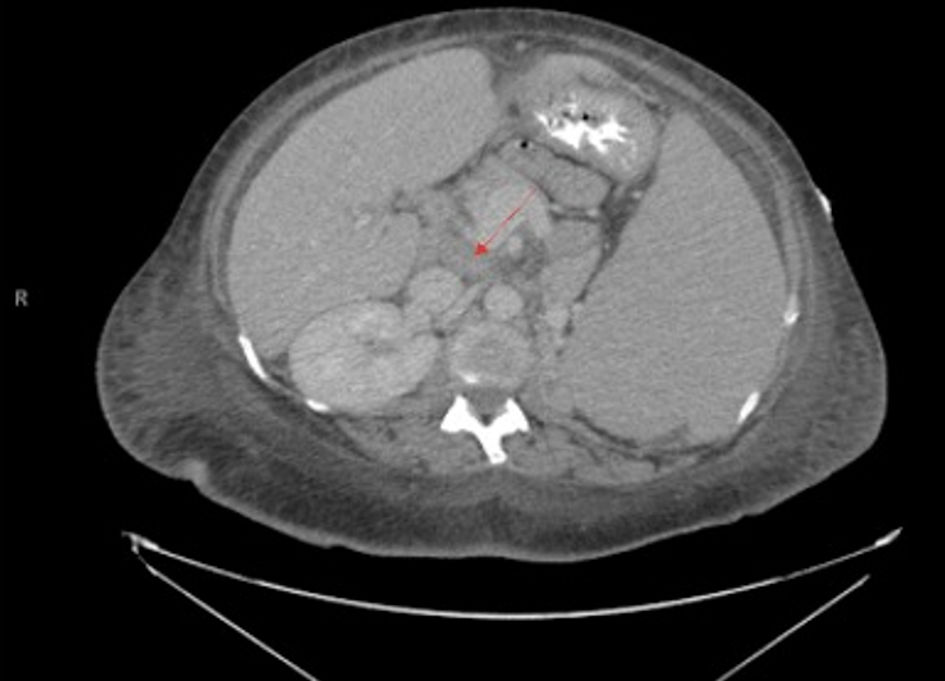
↓ Figure 2. Transverse view of CT
chest/abdomen/pelvis showing abdominal lymphadenopathy with noted splenomegaly, with arrow pointing to
prominent lymph node. A 3.4 × 2.1 cm nodule on the left adrenal gland is also noted.
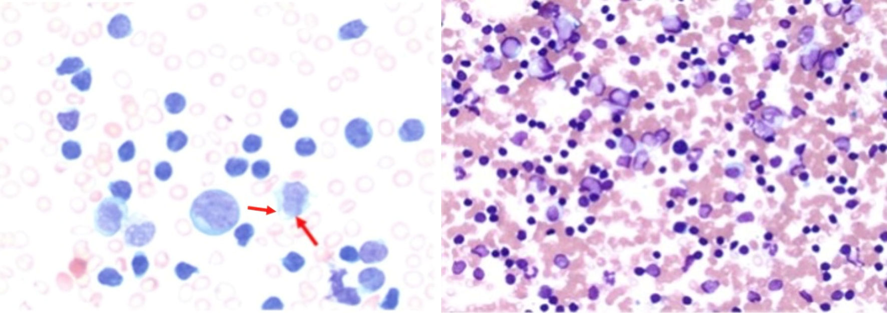
↓ Figure 3. Bone marrow aspirate demonstrating
atypical lymphoid cells with an increased nuclear-to-cytoplasmic ratio, irregular nuclear contours, and
condensed chromatin (arrows). Cytoplasmic blebs characteristic of leukemic prolymphocytes are present.
The bone marrow is nearly completely replaced by atypical lymphoid cells (magnification: × 50
(left) and × 10 (right)).