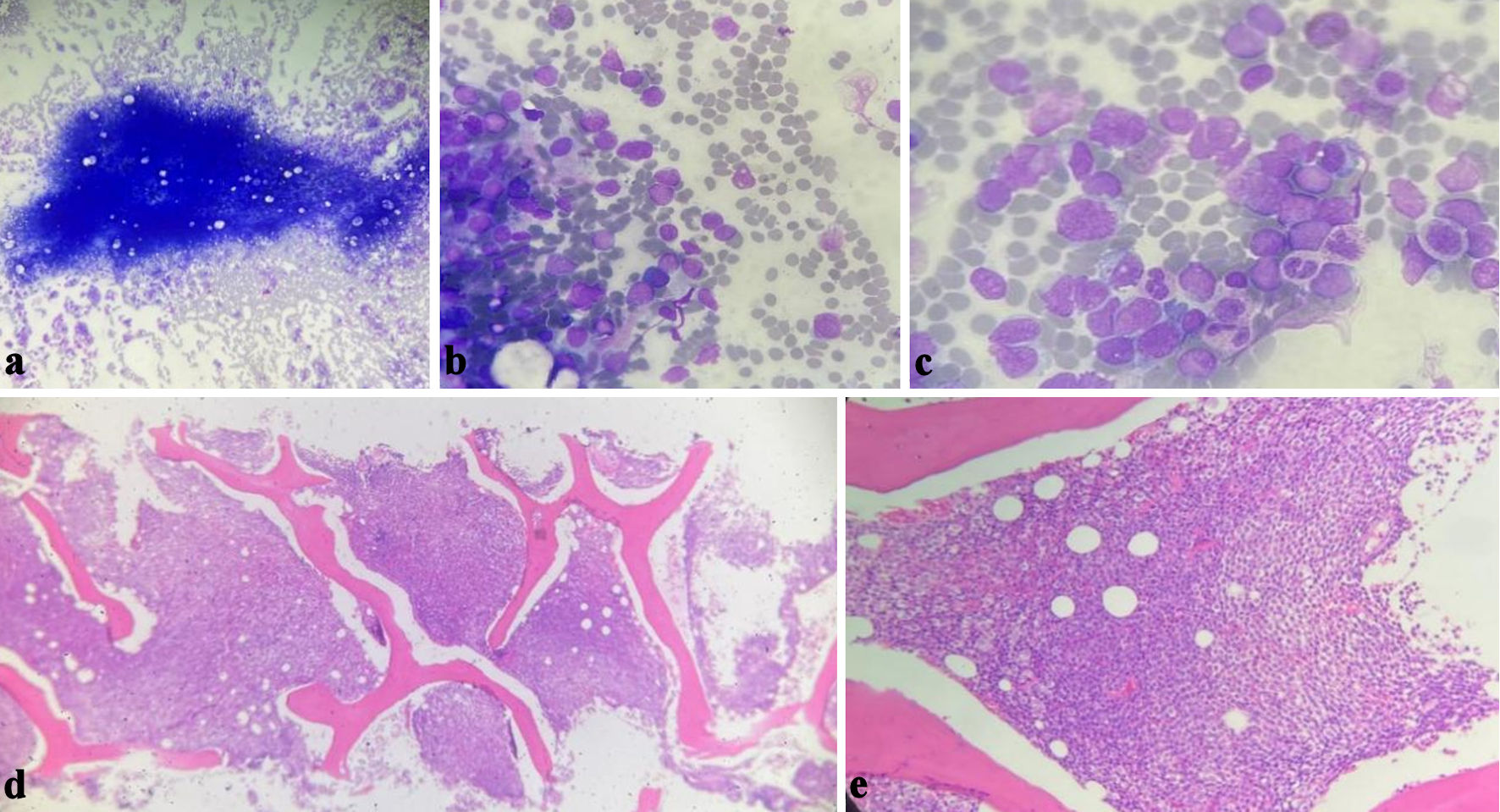
↓ Figure 1. Bone marrow aspirate (a, b, c)
showing the blasts infiltrating the bone marrow, the morphology is more toward myeloid leukemia. Bone
marrow biopsy (d, e) showing diffuse total infiltration by blasts.
| Journal of Hematology, ISSN 1927-1212 print, 1927-1220 online, Open Access |
| Article copyright, the authors; Journal compilation copyright, J Hematol and Elmer Press Inc |
| Journal website https://jh.elmerpub.com |
Case Report
Volume 14, Number 5, October 2025, pages 267-272
Acute Undifferentiated Leukemia Presented With Mediastinal Sarcoma
Figures
Table
| Time point | Event | Description |
|---|---|---|
| SOB: shortness of breath; CXR: chest X-ray; CT: computed tomography; PET: positron emission tomography; AUL: acute undifferentiated leukemia. | ||
| Day 0 | Patient presented with SOB | CXR showed right pleural effusion |
| Day 5 | Pleurocentesis | Exudative fluid |
| Day 8 | Echocardiogram | Pericardial effusion |
| Day 15 | Chest CT scan | Posterior mediastinal mass |
| Day 18 | Mediastinal mass biopsy | Myeloid sarcoma |
| Day 19 | PET scan | Bulky hypermetabolic mediastinal mass with bone marrow hyperactivity |
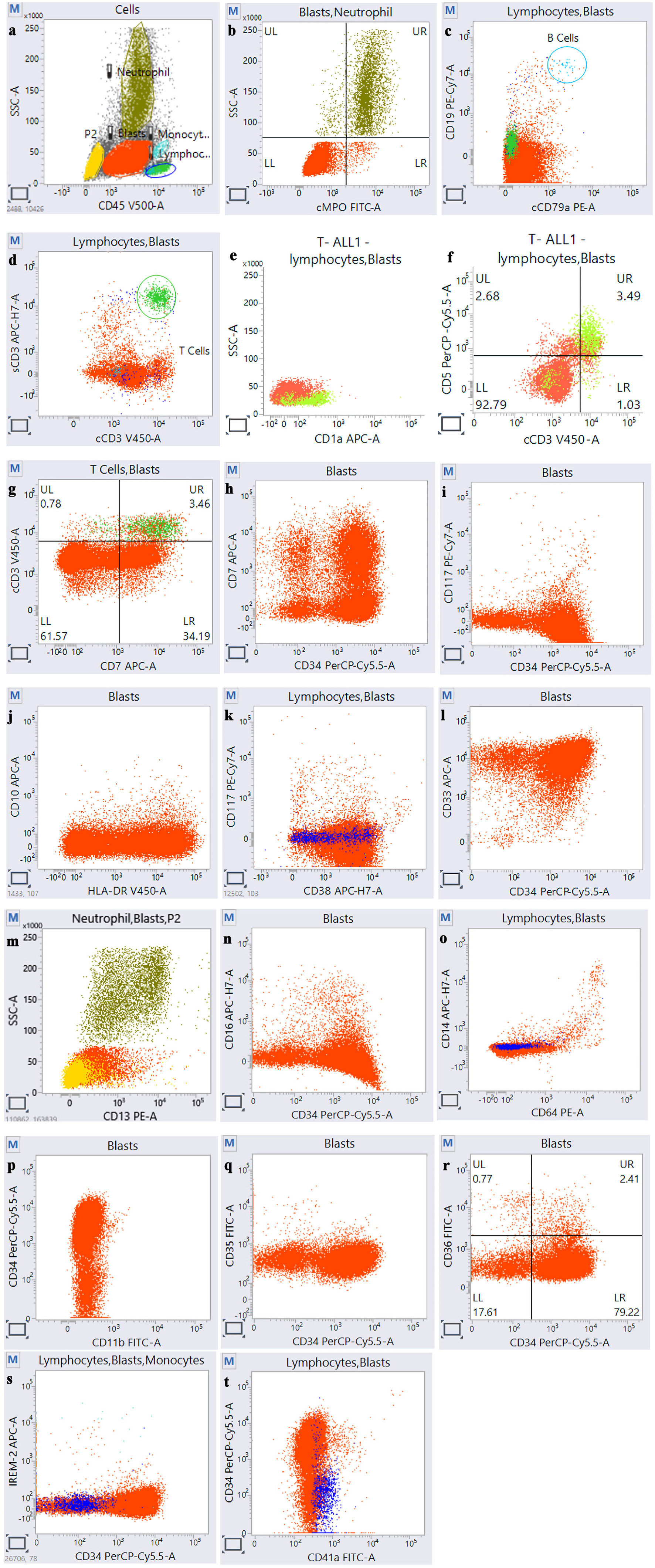
| Day 28 | Bone marrow aspirate | AUL |