Journal of Hematology, ISSN 1927-1212 print, 1927-1220 online, Open Access
Article copyright, the authors; Journal compilation copyright, J Hematol and Elmer Press Inc
Journal website https://jh.elmerpub.com

Case Report

Volume 14, Number 3, June 2025, pages 146-151


Ruxolitinib Is an Effective Therapy for Ciltacabtagene Autoleucel-Associated Parkinsonism in Multiple Myeloma

Figures

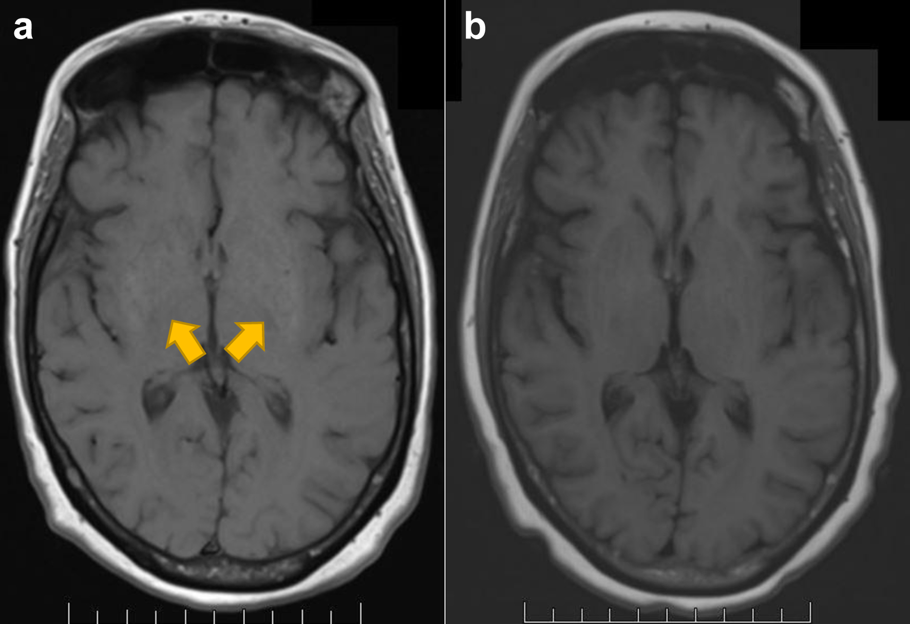
↓  Figure 1. Structural imaging with MRI T1WI of the brain for case 1. (a) Increased T1 signal of the basal ganglia (arrowheads) at diagnosis, not seen before cilta-cell infusion. (b) Post-treatment scan with resolution of prior T1 hyperintense signal of basal ganglia. MRI: magnetic resonance imaging; T1WI: T1-weighted image.
Figure 1.
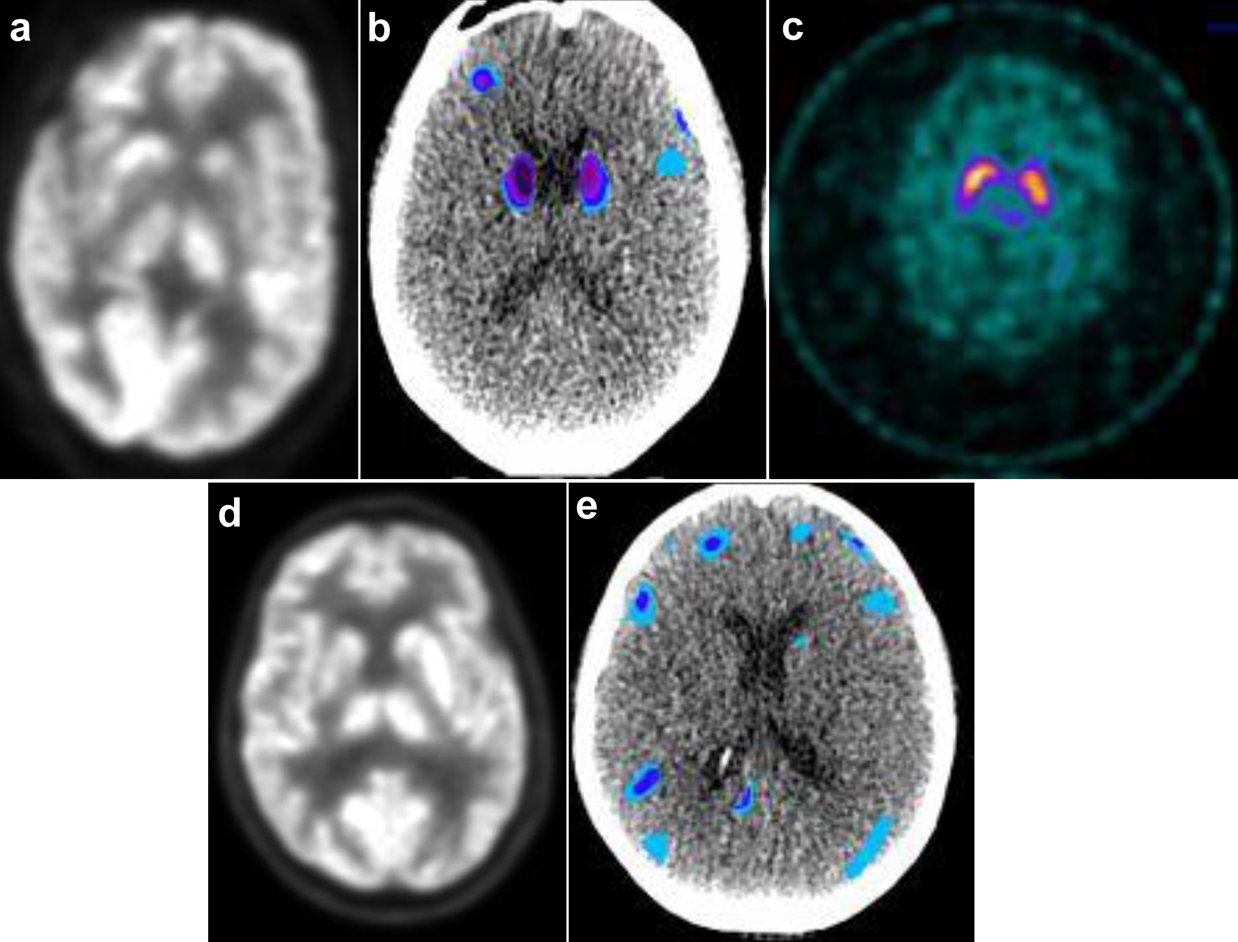
↓  Figure 2. Functional imaging for case 1 (a-c) and case 2 (d, e). (a) PET AC and (b) voxel-based analysis of brain PET normalized to standard dataset using MIMneuro software show significant decreased metabolism in bilateral caudate and frontal lobe/gyri. (c) DAT SPECT scan demonstrates symmetric normal uptake of basal ganglia. (d) PET AC and (e) normalized voxel-based analysis of PET brain demonstrate significant decreased metabolism in bilateral frontal lobe/gyri. AC: attenuation correction; DAT: dopamine activated transporter; PET: positron emission tomography; SPECT: single photon emission computed tomography.
Figure 2.

Table

↓  Table 1. Inflammatory Markers Before and After Ruxolitinib
 
Inflammatory markers Case 1 Case 2
sIL-2R: soluble interleukin-2 receptor.
At onset of parkinsonism before ruxolitinib
  Absolute lymphocyte count (1.1 - 3.3 × 10⁹/L) 1.4 × 10⁹ 7.6 × 10⁹
  Ferritin (16 - 150 ng/mL) > 30,000 9,258
  Fibrinogen (200 - 450 mg/dL) 90 99
  sIL-2R (223 - 770 U/mL) 8,180 2,826
After resolution of parkinsonism with ruxolitinib
  Absolute lymphocyte count (1.1 - 3.3 × 10⁹ /L) 1.1 × 10⁹ 0.7 × 10⁹
  Ferritin (16 - 150 ng/mL) 782 798
  Fibrinogen (200 - 450 mg/dL) 360 338
  sIL-2R (223 - 770 U/mL) 245 296