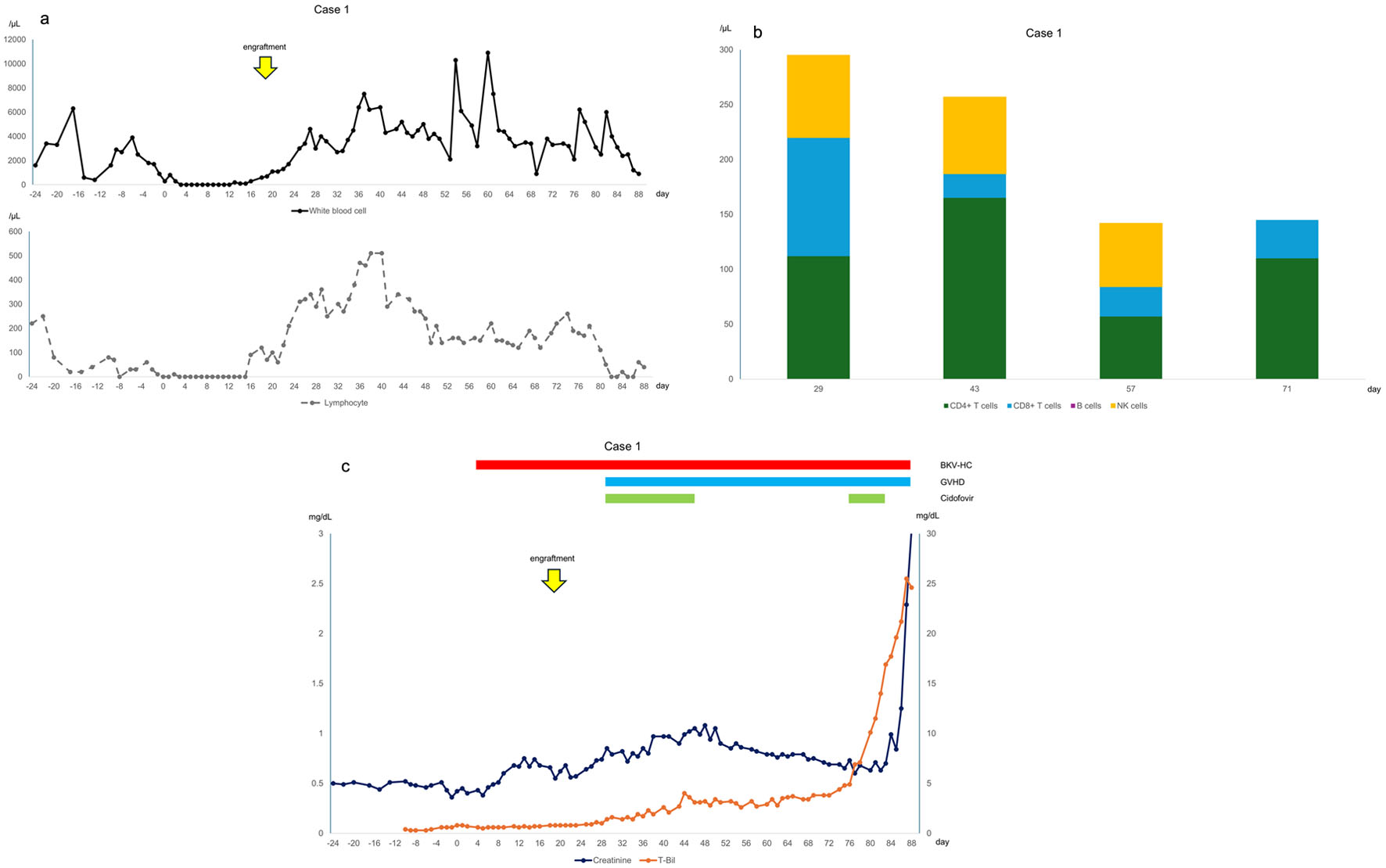
↓ Figure 1. (a) Changes in white blood cell and
lymphocyte counts, with day 0 representing the day of infusion. Lymphocyte counts decreased after day 50
when acute graft-versus-host disease (GVHD) worsened again. (b) Lymphocyte subsets, where
CD4+ T cells are CD3+CD4+CD8, CD8+ T cells are
CD3+CD4CD8+, B cells are CD19+CD20+, and natural killer
cells are CD3CD16+CD56+/CD3CD16CD56+. CD4+ T cells were
particularly low on day 57 when acute GVHD worsened. (c) Clinical course of the case, with day 0
indicating the day of infusion. BKV-HC: BK virus-associated hemorrhagic cystitis.
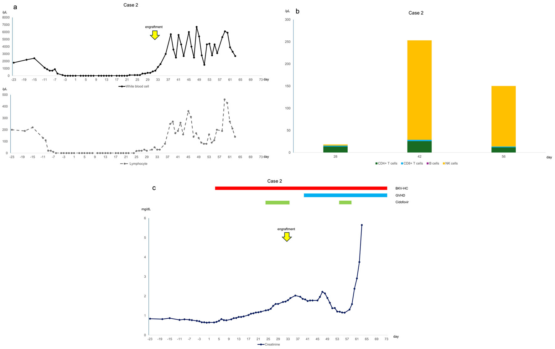
↓ Figure 2. (a) Changes in white blood cell and
lymphocyte counts, with day 0 representing the day of infusion. Lymphocyte counts were lower than those
in case 1. (b) Lymphocyte subsets. The absolute numbers of CD4+ T cells and CD8+ T
cells were lower than those in case 1. (c) Clinical course of the case, with day 0 representing the day
of infusion. Creatinine levels increased with the worsening of acute GVHD on day 40.
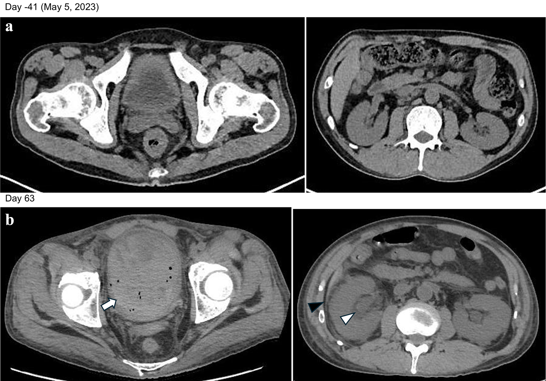
↓ Figure 3. (a) Findings after the completion of
three courses of Pola-BR. No abnormal findings were observed in the kidneys. (b) Heterogeneous
high-intensity areas in the bladder, indicating hemorrhage and blood clots (arrow). Dilation of the
renal pelvis (white arrowhead) and increased density of perirenal fat tissue (black arrowhead) were
observed. These findings indicate that the BK virus infection had spread from the bladder to the
kidneys.