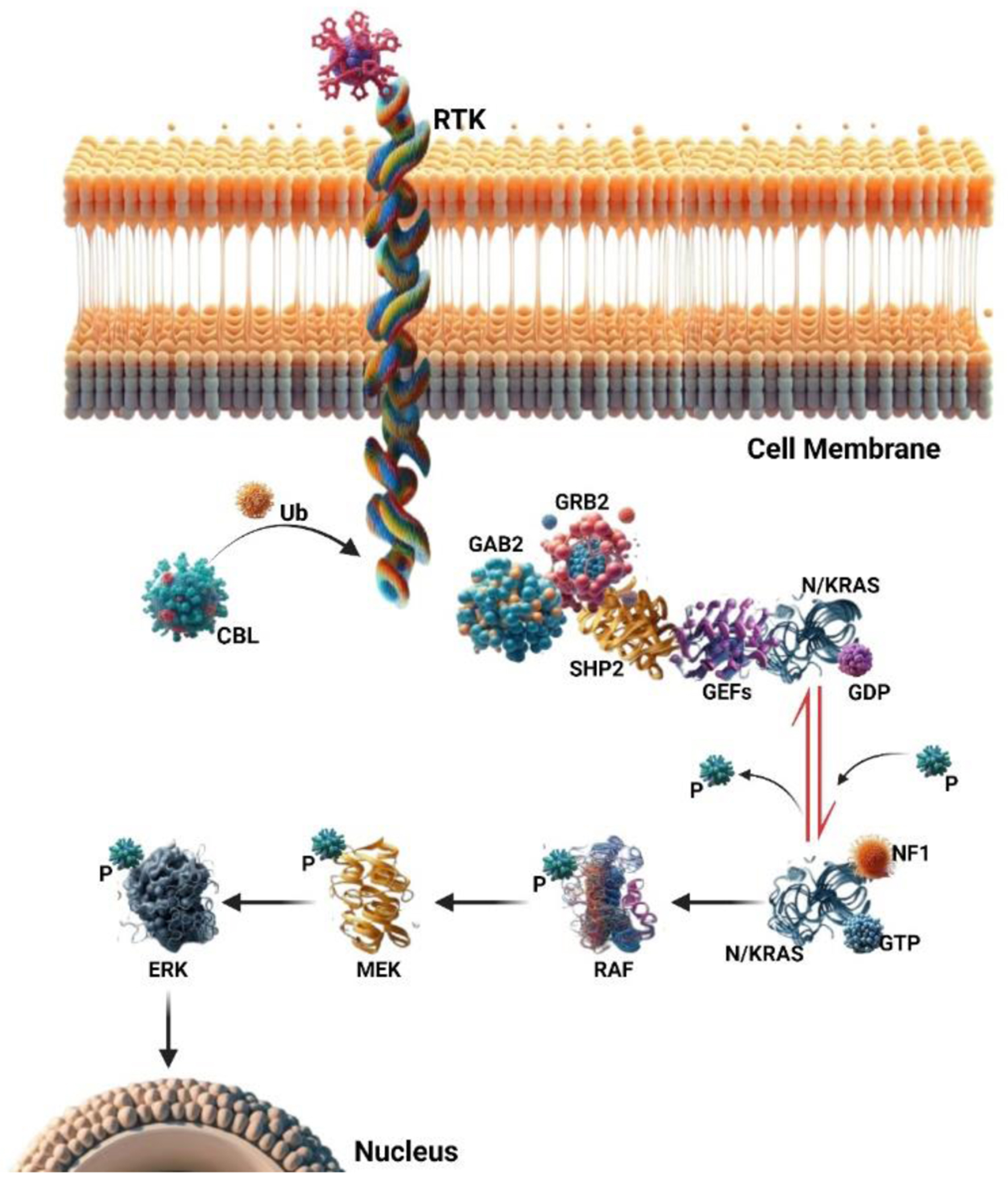
Journal of Hematology, ISSN 1927-1212 print, 1927-1220 online, Open Access
Article copyright, the authors; Journal compilation copyright, J Hematol and Elmer Press Inc
Journal website https://jh.elmerpub.com

Review

Volume 14, Number 2, April 2025, pages 43-55


MicroRNA Signatures: Illuminating Minimal Residual Disease Monitoring in Juvenile Myelomonocytic Leukemia - A Review

Figures

Figure 1.
Figure 1. The RAS signaling pathway involves small GTPase switch proteins, NRAS, and KRAS. These proteins act downstream of receptor and non-receptor tyrosine kinases (RTKs and TKs). RAS activation status is regulated through phosphorylation and dephosphorylation changes. Guanine nucleotide-exchange factors (GEFs) and GTPase activating proteins (GAPs) play opposing roles in this regulation. Upon RTK or TK stimulation, adaptor proteins (such as GAB2, GRB2, and SHP2) recruit GEFs, leading to RAS-GDP phosphorylation and activation of RAS-GTP. Active RAS then triggers a signaling cascade, sequentially activating RAF, MEK, and ERK proteins. These downstream signals ultimately control cell functions like proliferation, survival, and differentiation. GAPs (e.g., NF1) inactivate the RAS pathway by promoting RAS-GTP dephosphorylation to an inactive RAS-GDP form. Additionally, the ubiquitin ligase CBL negatively regulates the RAS pathway by targeting active RTKs for proteasomal degradation.
Figure 2.
Figure 2. Schematic diagram of microRNA (miRNA) biogenesis and function pathway.
Figure 3.
Figure 3. The use of microRNAs (miRNAs) as biomarkers in leukemia, employing both invasive and non-invasive methods for expression analysis. Non-invasive detection involves circulating miRNAs in body fluids like serum, blood, urine, and saliva. Invasive approaches extract intracellular miRNAs from bone marrow and peripheral blood cells. Extracellular miRNAs are also found in protein complexes, high-density lipoproteins, exosomes, and apoptotic bodies. These miRNAs are analyzed using advanced techniques such as microarrays, next generation sequencing (NGS), and quantitative reverse transcription PCR (qRT-PCR), showcasing their potential in leukemia diagnosis and monitoring.

Table

Table 1. A Comparative Overview of Traditional MRD Monitoring Methods vs. miRNA Signatures in JMML
 
AspectTraditional MRD monitoring methodsMiRNA signatures
ASO-RQ-PCR: allele-specific oligonucleotide-real-time quantitative PCR; EMT: epithelial-mesenchymal transition; Ig: immunoglobulin; JMML: juvenile myelomonocytic leukemia; MFC: multicolor flow cytometry; miRNA: microRNA; MRD: minimal residual disease; NGS: next-generation sequencing.
MethodologyPCR-based strategies: e.g., ASO-RQ-PCR. Immunophenotype-based strategies: MFC for immunophenotypingNGS-based clonality assays: high-throughput sequencing of Ig rearrangements provides ultrasensitive MRD detection.
SensitivityModerate sensitivity (approximately 1 × 10⁻⁵)Ultra-sensitive detection, capable of identifying very low levels of residual leukemia cells
SpecificityProvides moderate specificity through detection of immunophenotypic markersIdentifies specific miRNA patterns associated with distinct cancer subtypes, grades, and stages
Sample typeTypically requires invasive procedures (e.g., bone marrow aspirates)Non-invasive approach using easily accessible body fluids (blood, urine)
Clinical utilityWell established in clinical practice for various hematological malignanciesShows promise for early diagnosis and improved prognosis, particularly in breast cancer
CustomizationLimited to standardized assays with fixed parametersCan be tailored to individual patient profiles based on unique miRNA expression patterns
Functional insightsFocuses on identifying specific immunophenotypic markers without broader pathway insightsProvides additional information on miRNA networks and their roles in oncogenic processes, such as EMT and cellular plasticity
Role in EMTNot directly linked to EMTDirectly implicated in EMT and cellular plasticity, playing a role in tumorigenesis
Early detectionMay not detect MRD at very early stages due to limited sensitivityCapable of detecting MRD at earlier stages, offering a potential advantage in timely intervention
Cost and speedCost and turnaround time can vary; some methods may be both time-consuming and expensive.Emerging evidence suggests potential for a more cost-effective and rapid detection method, though further validation is ongoing.
Recommended sensitivity thresholdGenerally, a threshold of at least 10-4 is required for reliable assessment.Studies suggest that thresholds as low as 10-6 might correlate with prolonged progression-free survival, warranting further research and validation.