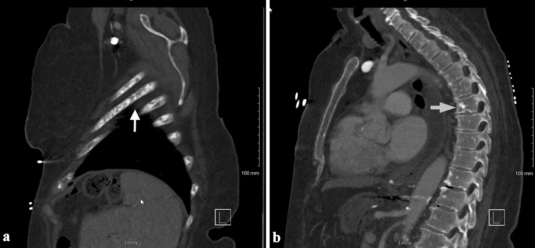
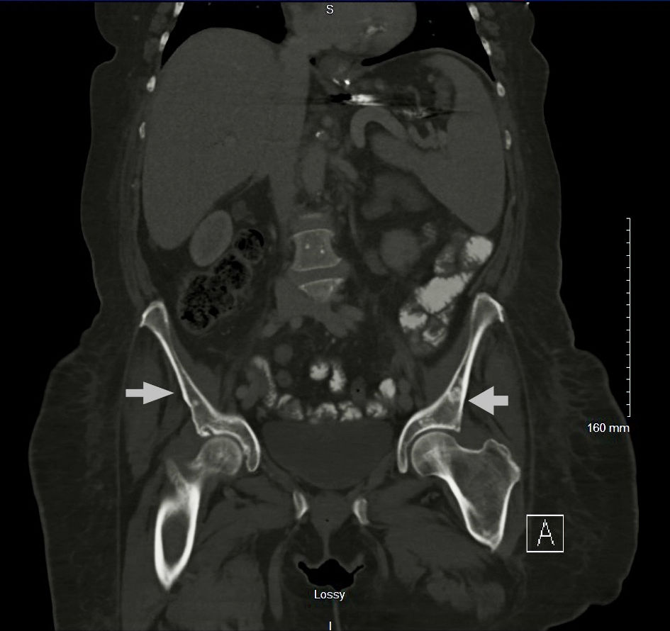
↓ Figure 1. CT chest with contrast showing the
osteoblastic lesions in the ribs (a) and vertebrae (b) (arrows). CT: computed tomography.
| Journal of Hematology, ISSN 1927-1212 print, 1927-1220 online, Open Access |
| Article copyright, the authors; Journal compilation copyright, J Hematol and Elmer Press Inc |
| Journal website https://jh.elmerpub.com |
Case Report
Volume 14, Number 1, February 2025, pages 32-37
A Rare Case of Acute Aleukemic Mast Cell Leukemia With Osteoblastic Lesions in the Appendicular Skeleton
Figures

Table
| Laboratory test | Day 1 | Reference range |
|---|---|---|
| AST: aspartate aminotransferase; ALT: alanine aminotransferase; BUN: blood urea nitrogen. | ||
| White blood count (WBC) | 3.3 | 4 - 11 × 103/µL |
| Hemoglobin | 11.3 | 12 - 16 g/dL |
| Hematocrit | 32 | 36-48% |
| Platelet | 104 | 150 - 450 × 103/µL |
| Alkaline phosphatase | 362 | 40 - 129 U/L |
| Total bilirubin | 2 | 0.0 - 1.0 mg/dL |
| AST/ALT | 38/23 | 10 - 40 U/L |
| Potassium | 2.5 | 3.5 - 5 mmol/L |
| BUN | 12 | 8 - 23 mg/dL |
| Creatinine | 0.57 | 0.5 - 1.0 mg/dL |
| Total protein | 5.9 | 6.0 - 8.3 gm/dL |
| Albumin | 3.1 | 3.5 - 5.2 gm/dL |
| Calcium | 8.6 | 8.5 - 10.2 mg/dL |