Journal of Hematology, ISSN 1927-1212 print, 1927-1220 online, Open Access
Article copyright, the authors; Journal compilation copyright, J Hematol and Elmer Press Inc
Journal website https://jh.elmerpub.com

Original Article

Volume 14, Number 1, February 2025, pages 1-13


Prevalence of Viral Infections and Serious Complications in Pediatric Hematopoietic Stem Cell Transplant Patients: A Ten-Year Single-Institution Retrospective Study

Figures

↓  Figure 1. Distribution of infectious episodes by virus, along post-transplant phases (a), graft sources (b), age groups (c); and pie charts of distribution of viral infection episodes by age groups or graft sources (d). ADNV: adenovirus; BK virus: human polyoma virus 1; BM: bone marrow; CMV: cytomegalovirus; EBV: Epstein-Barr virus; GvHD: graft-versus-host disease; HHV6: human herpes virus 6; HSV: herpes simplex virus; PBSC: peripheral blood stem cell; pHSCT: pediatric hematopoietic stem cell transplant; UCB: umbilical cord blood; VZV: varicella-zoster virus.
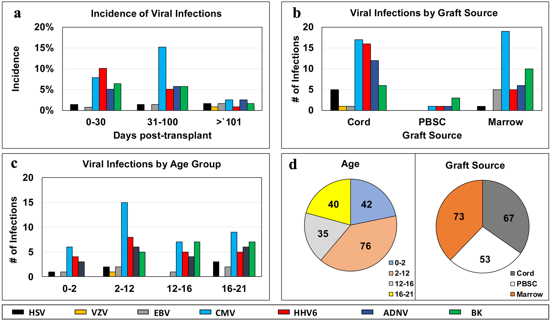
Figure 1.
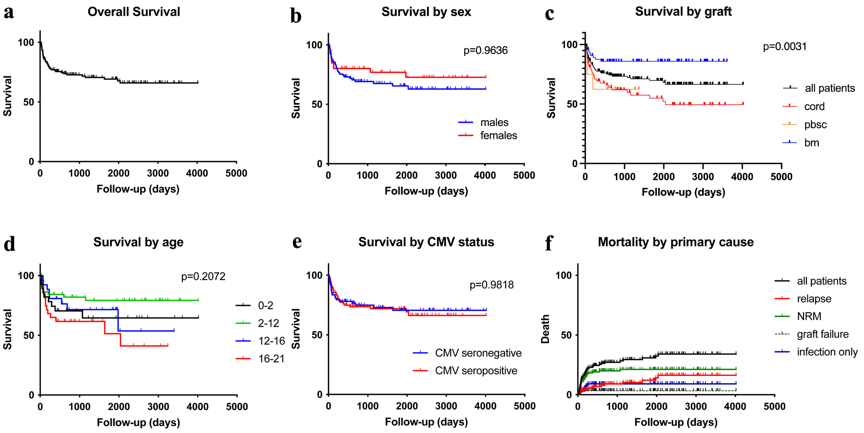
↓  Figure 2. Kaplan-Meier plots for the study population: overall survival (a), overall survival by sex (b), overall survival by graft sources (c), overall survival by age groups (d), overall survival by patient’s CMV status (e), and mortality by primary cause of death (f). CMV: cytomegalovirus.
Figure 2.

Tables

↓  Table 1. Demographics of Study Patients
 
Demographic variables Age P-value
0 - 2 years old (n = 30) 2 - 12 years old (n = 58)a 12 - 16 years old (n = 29)b 16 - 21 years old (n = 34)
Sex 0.1018
  Female 12 22 4 12
  Male 18 36 25 22
Ethnicity 0.6918
  Hispanic 19 36 20 21
  White 6 15 5 8
  Middle Eastern 4 6 2 2
  Asian 1 1 1 3
  NOS 0 0 1 0
Primary disease 0.0093
  Malignant 13 37 21 28
  Non-malignant 17 21 8 6
Disease classification 3.6835 × 10⁻⁵
  Acute leukemia 9 35 13 24
  MDS/MPL 1 1 0 0
  Primary immunodeficiency 11 5 0 1
  IDM 0 1 1 0
  Histiocytic disorder 3 2 1 0
  Chronic leukemia 0 6 6 5
  Bone marrow failure 2 6 1 0
  Hemoglobinopathy 4 2 6 1
  PTLD 0 0 1 0
  Lymphoma 0 1 0 3
  Solid tumor 0 0 1 0
Disease status 0.2248
  CR1 9 17 12 16
  CR1/MRD+ 1 2 0 0
  CR2 1 15 6 11
  CR2/MRD+ 0 1 0 0
  CR3 1 3 1 1
  CR4 0 2 0 0
  PR 3 2 0 0
  Aplastic 0 1 0 0
  Active disease 1 0 1 1
  N/A 14 15 9 5
Cell source 0.03260
  Cord 18 31 9 9
  PBSC 0 5 2 4
  Marrow 12 22 18 21
0 - 2 years old (n = 42) 2 - 12 years old (n = 76)a 12 - 16 years old (n = 35)b 16 - 21 years old (n = 40)
A total of 151 patients were distributed across four age groups. aOne patient was originally diagnosed and treated for juvenile myelomonocytic leukemia and later developed post-transplant lymphoproliferative disorder (PTLD). bOne patient was diagnosed and treated for histiocytic disorder and later developed chronic leukemia. Sex and ethnicity were self-reported, patients who declined to report were documented as not otherwise specified (NOS). Malignant diseases included acute leukemia, myelodysplastic syndrome/myeloproliferative disorder (MDS/MPL), chronic leukemia, lymphoma, and solid tumors. Non-malignant diseases included primary immunodeficiency, inborn disease of metabolism (IDM), histiocytic disorders, bone marrow failure, hemoglobinopathy, and platelet and other inherited disorders (PLD). With regard to disease status, complete response (CR) refers to no detectable evidence of malignancy on imaging and/or tissue samples. Partial response (PR) refers to a significant reduction in tumor burden, with minimal disease detectable on imaging and/or tissue samples. Mean residual disease positive (MRD+) refers to detectable disease based on molecular and/or tissue sampling. Both donor and recipient serologies were documented for CMV and HSV. GCV prophylaxis distribution described for the 81 recipient CMV+ cases. Rates of ganciclovir and acyclovir prophylaxis were documented in only the recipient seropositive CMV and HSV patient groups. P-values of χ2 test are shown. CMV: cytomegalovirus; GCV: ganciclovir; HRD: haplo-related donor; HSV: herpes simplex virus; MRD: matched related donor; MUD: matched unrelated donor; PBSC: peripheral blood stem cell; TBI: total body irradiation.
Donor source 0.1809
  MUD 24 37 18 16
  MRD 6 17 8 15
  HRD 0 4 3 3
Conditioning 0.0422
  Conditioning, no TBI 24 31 16 16
  TBI conditioning 6 27 13 18
CMV serology
  Donor 0.2946
    CMV- 12 28 17 12
    CMV+ 17 30 12 22
    Missing 1 0 0 0
  Recipient 0.2473
    CMV- 12 28 17 12
    CMV+ 17 30 12 22
    Equivocal 1 0 0 0
GCV prophylaxis 0.7643
  Yes 13 24 11 18
  No 4 6 1 4
HSV serology 0.3152
  HSV- 16 26 18 21
  HSV+ 14 32 11 13
VZV serology 0.7567
  VZV+ 3 14 5 6
  VZV- 3 3 3 3
  Not tested 24 41 21 25
Acyclovir prophylaxis 0.5516
  Yes 17 35 21 23
  No 13 23 8 11


↓  Table 2. CMV Viral Infections in the Post-Transplant Period
 
CMV serology Infectious event (days post-transplant)
No. of infections 0 - 30 31 - 100 101 - 730 CMV disease
Patients were organized into four recipient (R) and donor (D) serology groups, with either seropositive or seronegative CMV results (+ or -). Incidence of CMV infection was divided into three post-transplant time periods (0 - 30, 31 - 100, and 101 - 730 days). Incidence of CMV disease in these recipient/donor serology groups was also documented. Three patients were excluded from this analysis, as they were missing CMV serology data or had equivocal serology result. P-value = 0.00192 across all groups. P-value = 0.6320 between R+/D- infection and R+/D- infection. P-value = 0.2875 between R+/D- and R+/D- CMV disease. CMV: cytomegalovirus.
R-/D- (n = 31) 0 0 0 0 0
R-/D+ (n = 37) 3 1 2 0 0
R+/D- (n = 30) 12 6 5 1 7
R+/D+ (n = 50) 22 5 15 2 7
Total 37 12 22 3 14


↓  Table 3. HHV6 Infections in the Post-Transplant Period
 
HHV6 infections Infectious event (days post-transplant)
No. of infections 0 - 30 31 - 100 101 - 730 Encephalitis
Patients were grouped by cell source. Incidence of HHV6 divided into three post-transplant time periods (0 - 30, 31 - 100, and 101 - 730 days). Rates of all viral encephalitis (including HSV, VZV, HHV6, and ADNV) were also organized by cell source. P-value = 0.01479 for graft source and number of infections. ADNV: adenovirus; HHV6: human herpes virus 6; HSV: herpes simplex virus; PBSC: peripheral blood stem cell; VZV: varicella-zoster virus.
Cord 16 11 4 1 5
PBSC 0 0 0 0 0
Marrow 5 2 3 0 0
Total 21 13 7 1 5


↓  Table 4. Descriptions and Outcomes of Patients Diagnosed With Viral Encephalitis
 
Patient (age in years) Primary diagnosis Cell source Time from transplant (days) Comorbidities Outcomes (treatment/status/cause of death)
The characteristics, complications, and time course of eight patients diagnosed with viral encephalitis were described. The primary diagnosis details the disease indicated for HSCT. Time from transplant to diagnosis of encephalitis was categorized into three broad post-transplant periods. Common comorbidities of HSCT therapy, as well as disease-specific complications were also documented. The antiviral therapies used to treat encephalitis included acyclovir, ganciclovir, foscarnet, and cidofovir. Patient status was detailed as alive, recovered from encephalitis, deceased from encephalitis (E+ denoting number of days survived with encephalitis), and overall deceased date in the post-transplant period (D+ denoting number of days survived from transplant). Major causes of death in addition to encephalitis were also detailed. aOne patient was lost to follow-up and was confirmed to be deceased by an outside hospital. ADNV: adenovirus; AKI: acute kidney injury; ALL: acute lymphocytic leukemia; AML: acute myeloid leukemia; AMS: altered mental status; ARF: acute renal failure; BS: bacterial sepsis; CMV: cytomegalovirus; FS: fungal sepsis; GF: graft failure; GvHD: graft-versus-host disease; HHV6: human herpes virus 6; HSCT: hematopoietic stem cell transplant; HSV: herpes simplex virus; LTFU: lost to follow-up; UCB: umbilical cord blood; UTI: urinary tract infection; VRE: vancomycin-resistant enterococcus; VZV: varicella-zoster virus.
E1 (18) ALL/HHV6 UCB 0 - 30 BK hemorrhagic cystitis, bacterial sepsis, fungal sepsis Foscarnet, ganciclovir; deceased E+ 139; D+ 166; BS, FS, HHV6 encephalitis, BK
E2 (5) AML/HHV6 UCB 0 - 30 HHV6 viremia, GvHD, ADNV enteritis Foscarnet, ganciclovir; recovered from encephalitis; deceased D + 602; relapsed disease, BS.
E3 (20) AML/ADNV UCB 31 - 100 HHV6 viremia, GvHD, ADNV enteritis, VRE UTI Cidofovir; deceased E+ 43; D+ 70; AMS, AKI, GvHD, HHV6
E4 (4) AML/HHV6 UCB 0 - 30 HHV6 viremia, BK hemorrhagic cystitis, Klebsiella UTI Ganciclovir; recovered from encephalitis; alive
E5 (8) ALL/HHV6a UCB 0 - 30 ADNV enteritis, HSV viremia, Aspergillus pneumonia, C. diff colitis Acyclovir; deceased E+ 39; D+ 64; FS, HHV6, ADNV, ARF
E6 (9) AML/HHV6 UCB 0 - 30 Graft failure, fungal sepsis, HHV6, ADNV Foscarnet, ganciclovir; deceased E+ 17; D+ 39; GF, FS, HHV6, ADNV, ARF
E7 (15) AML/VZVa UCB 101 - 730 GvHD, CMV viremia, BK viremia, fungal sepsis, ADNV enteritis Acyclovir; deceased date unknown; GF, LTFU