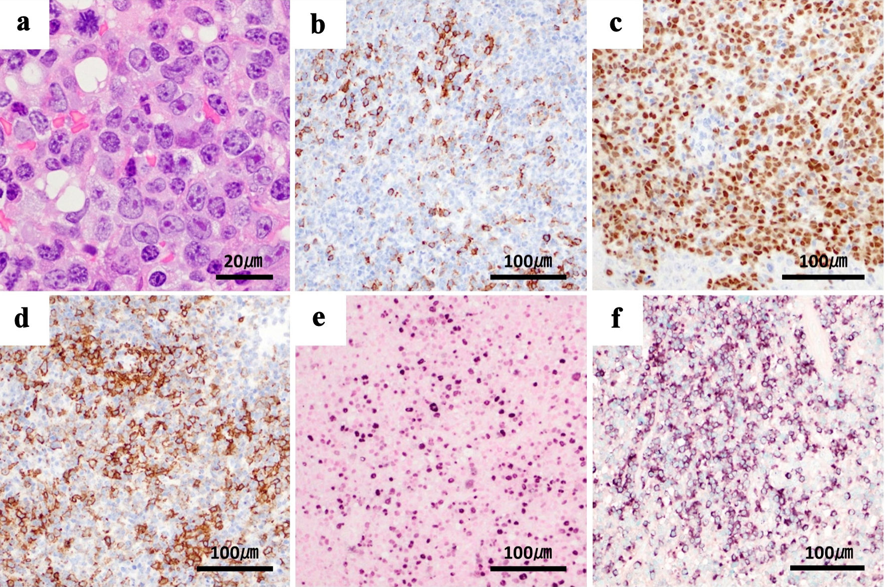
Journal of Hematology, ISSN 1927-1212 print, 1927-1220 online, Open Access
Article copyright, the authors; Journal compilation copyright, J Hematol and Elmer Press Inc
Journal website https://jh.elmerpub.com

Case Report

Volume 13, Number 6, December 2024, pages 290-294


Spontaneous Regression of Plasmablastic Lymphoma Associated With Methotrexate After Withdrawal

Figures

Figure 1.
Figure 1. Biopsy of the oral ulcer shows tumor cells with plasmablastic differentiation. Tumor cells show immunoreactivity for plasma cell-associated antigen MUM-1 and partial reactivity for CD138, variable positivity for B-cell markers CD20 and CD79a, and cytoplasmic λ light chain restriction. EBV is present, and MIB-1 is positive in over 90% of cells. These findings are compatible with plasmablastic lymphoma. (a) Tumor cells in oral ulcer (hematoxylin and eosin staining), × 100. (b) Immunoblastic cells exhibiting immunoreactivity with CD138. (c) MUM-1. (d) CD20. (e) Nuclear positivity with EBER in situ hybridization. (f) Cytoplasmic λ light chain restriction, × 20. MUM-1: multiple myeloma oncogene 1; CD: cluster of differentiation; EBV: Epstein-Barr virus; EBER: EBV-encoded small RNA; RNA: ribonucleic acid; MIB-1: monoclonal antibody Ki-67.
Figure 2.
Figure 2. (a) PET-CT shows FDG accumulation in the known right mandibular tumor. (b) Multiple mass-like shadows are observed in the lung fields, but no FDG accumulation is noted. PET-CT: positron emission tomography-computed tomography; FDG: 18F-fluorodeoxyglucose.
Figure 3.
Figure 3. Chest CT and oral ulcer findings. The oral ulcer lesions and multiple pulmonary nodules (arrows) reduced with only discontinuation of methotrexate. (a) At the time of initial diagnosis. (b) One month later. (c) Three months later. CT: computed tomography.