| Journal of Hematology, ISSN 1927-1212 print, 1927-1220 online, Open Access |
| Article copyright, the authors; Journal compilation copyright, J Hematol and Elmer Press Inc |
| Journal website https://jh.elmerpub.com |
Review
Volume 15, Number 5, October 2026, pages 225-235

Ferroptosis and Oxidative Stress in Sickle Cell Anemia
Neda Alvarez-Ortegaa, b, Victor Eljach Cordobac, Sheccid Genes Guerrac, Sonia M. Julio Huetoc, Jesus D. Rangel Pupoc, Jerson A. Zuniga Parra, Emilia M. Polo-Carrillob, Jenny Palomares-Bolanosa, b, d
aProject Management Center, University Institution Mayor of Cartagena, Cartagena de Indias, Colombia
bUniversity Institution Mayor of Cartagena, Cartagena de Indias, Colombia
cPharmaceutical Chemistry Program, School of Pharmaceutical Sciences, University of Cartagena, Cartagena de Indias, Colombia
dCorresponding Author: Jenny Palomares-Bolanos, Project Management Center, University Institution Mayor of Cartagena, Cartagena de Indias, Colombia
Manuscript submitted June 17, 2026, accepted August 13, 2026, published online October 31, 2026
Short title: Ferroptosis in Sickle Cell Anemia
doi: https://doi.org/10.14740/jh2225
| Abstract | ▴Top |
Ferroptosis, an iron-dependent cell death, plays a key role in the pathophysiology of sickle cell disease (SCD). This study is a narrative review on oxidative stress and ferroptosis in children with SCD in Africa and Colombia, through literature searches to select articles published between 2019 and 2024. The search included terms such as “sickle cell,” “oxidative stress,” “ferroptosis,” “Africa,” and “Colombia,” consulting databases like PubMed, Springer, Google Scholar and ScienceDirect, and selecting 11 relevant studies. The results show that ferroptosis, induced by the accumulation of free iron, lipid peroxidation, and increased reactive oxygen species (ROS), worsens complications such as kidney damage, vase-occlusive crises, and chronic inflammation. In Africa, factors like low hepcidin levels and genetic mutations, such as Q248H in ferroportin, increase oxidative stress and susceptibility to ferroptosis. In Colombia, the high prevalence of hemoglobin S in the Caribbean and Pacific regions is associated with severe crises and significant tissue damage. Clinical cases suggest that hydroxyurea reduces vaso-occlusive crises by raising fetal hemoglobin (HbF) levels and chelating free iron. Although ferroptosis is a secondary mechanism in the pathophysiology of SCD, it significantly amplifies complications, making it essential to implement therapeutic strategies that regulate iron homeostasis and reduce oxidative stress using antioxidants and lipid peroxidation inhibitors.
Keywords: Anemia; Oxidative Stress; Ferroptosis; Africa; ROS
| Introduction | ▴Top |
Hematological diseases are pathologies in which the blood and hematopoietic organs are affected. Among these are non-malignant hematological diseases, which include various conditions such as sickle cell diseases (SCD) [1]. These are a group of inherited disorders of the hemoglobin molecule characterized by chronic hemolytic anemia [2]. SCD is an autosomal recessive hemoglobin disorder resulting from homozygous inheritance or compound heterozygosity of a mutation in the β-globin gene. A single base-pair point mutation (GAG to GTG) results in the substitution of the amino acid glutamic acid (hydrophilic) with valine (hydrophobic) at the sixth position of the β chain of hemoglobin, giving rise to hemoglobin S (HbS) [3, 4]. This mutation leads to the production of abnormal β chains in hemoglobin, the protein that constitutes approximately 70% of the erythrocyte mass, promoting polymerization and deformation of the normal erythrocyte shape. This process results in the obstruction of small-diameter blood vessels [5], causing hypoxia in critical organs [6]. In addition, these cells are more prone to premature death.
SCD has had a high global prevalence and a substantial social impact, with significant mortality occurring in the first years of life. A high prevalence has been reported in sub-Saharan Africa, the Mediterranean region, the Middle East, and India [7]. In addition, in recent years, there has been an increase in the number of births of children with SCD in Latin America and the Caribbean, along with a decrease in Central Europe, Central Asia, North Africa, and the Middle East [8]. The number of individuals living with SCD has increased by 41.4% over the past 20 years, reaching 7,74 million in 2021. It is estimated that 300,000 newborns are diagnosed with SCD worldwide [9], with 75% of cases occurring in Africa [10]. Sickle cell anemia (SCA) is most prevalent in sub-Saharan Africa, with an incidence ranging from 1% to 5% in most populations. In Ghana, 2% of newborns are affected by SCA, of whom the most severe form (homozygous HbS or SCA) accounts for 55% [11].
Patients may experience a wide range of symptoms, including recurrent pain, chronic anemia, and multi-organ damage, leading to significant clinical morbidity and early mortality [12]. The burden of mortality in Africa remains disproportionately high compared to that of children without SCD. This disparity is partly attributable to the limited availability of healthcare resources and the implementation of less effective intervention measures for managing SCD [13]. The aim of this study is to analyze the role of ferroptosis as a pathogenic mechanism in SCA, with a particular focus on its relationship with oxidative stress and its impact on populations in Africa and Colombia. Through a systematic and bibliographic review, this study seeks to explore the connections between symptoms, clinical signs, triggering factors, and clinical outcomes related to the ferroptosis pathway. Furthermore, it aims to evaluate how this process may influence patients’ physical condition and contribute to the development of therapeutic strategies.
A narrative literature review was conducted by reviewing scientific articles and selecting case studies. The search was limited to the period between 2019 and 2024 and employed the terms “sickle cell,” “oxidative stress,” “child,” “ferroptosis,” “Africa,” and “Colombia.” These descriptors were applied to the title, abstract, and full text sections of four electronic databases: PubChem, ScienceDirect, Springer, and Google Scholar. Relevant or potentially relevant citations were identified using the Publish or Perish software, while duplicate records were removed using Covidence, as illustrated in Table 1 [14–23].
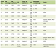 Click to view | Table 1. Selected Studies for Screening for SCA and Its Associated Complications |
This review included studies that met the following criteria; original research articles published between 2019 and 2024, based on research on SCD or SCA, and addressing oxidative stress, ferroptosis, iron metabolism, lipid peroxidation, or related molecular mechanisms. Studies were also required to include human participants with SCD or experimental models directly related to this disease, and to have been conducted in African countries or in Colombia, or to provide data relevant to these regions. In addition, observational studies (cross-sectional, case-control, and cohort studies), randomized clinical trials, experimental studies, and case reports documenting at least one patient with SCD were considered eligible. Articles published in English or Spanish were considered for this descriptive analysis.
On the other hand, review articles, systematic reviews, meta-analyses, editorials, letters to the editor, conference abstracts, undergraduate or graduate theses, book chapters, and studies not focused on SCD were excluded. Also excluded were studies that did not evaluate oxidative stress, ferroptosis, or related biological pathways; articles lacking sufficient methodological information or full-text access; duplicate publications; and studies conducted in populations unrelated to the objectives of this review.
After duplicate removal, titles and abstracts were screened according to the predefined inclusion and exclusion criteria. Potentially eligible studies underwent full-text assessment. Eligible articles were then categorized according to their primary focus on oxidative stress, ferroptosis, and SCD, with particular emphasis on studies conducted in African populations and Colombia. The second category consisted of excluded documents, such as articles unrelated to SCD, review articles, letters to the editor, commentaries, conference abstracts, and undergraduate theses. In addition, observational studies including cohort studies, case control studies, randomized controlled trials, and case reports that documented at least one case of SCD were reviewed.
| SCD | ▴Top |
This is an autosomal recessive disease that results from a mutation (dbSNP rs334(T)) in the gene encoding the β-globin chain of hemoglobin. This genetic alteration causes the substitution of glutamic acid by valine (Fig. 1) [24, 25], leading to the formation of aberrant hemoglobin (HbS), which polymerizes under low-oxygen conditions, thereby inducing erythrocyte hemolysis and episodes of vaso-occlusive crises [26, 27]. The pathophysiology of SCD involves chronic inflammation, oxidative stress, and vaso-occlusion, resulting in a wide range of clinical outcomes, including pain crises and progressive organ damage [28].
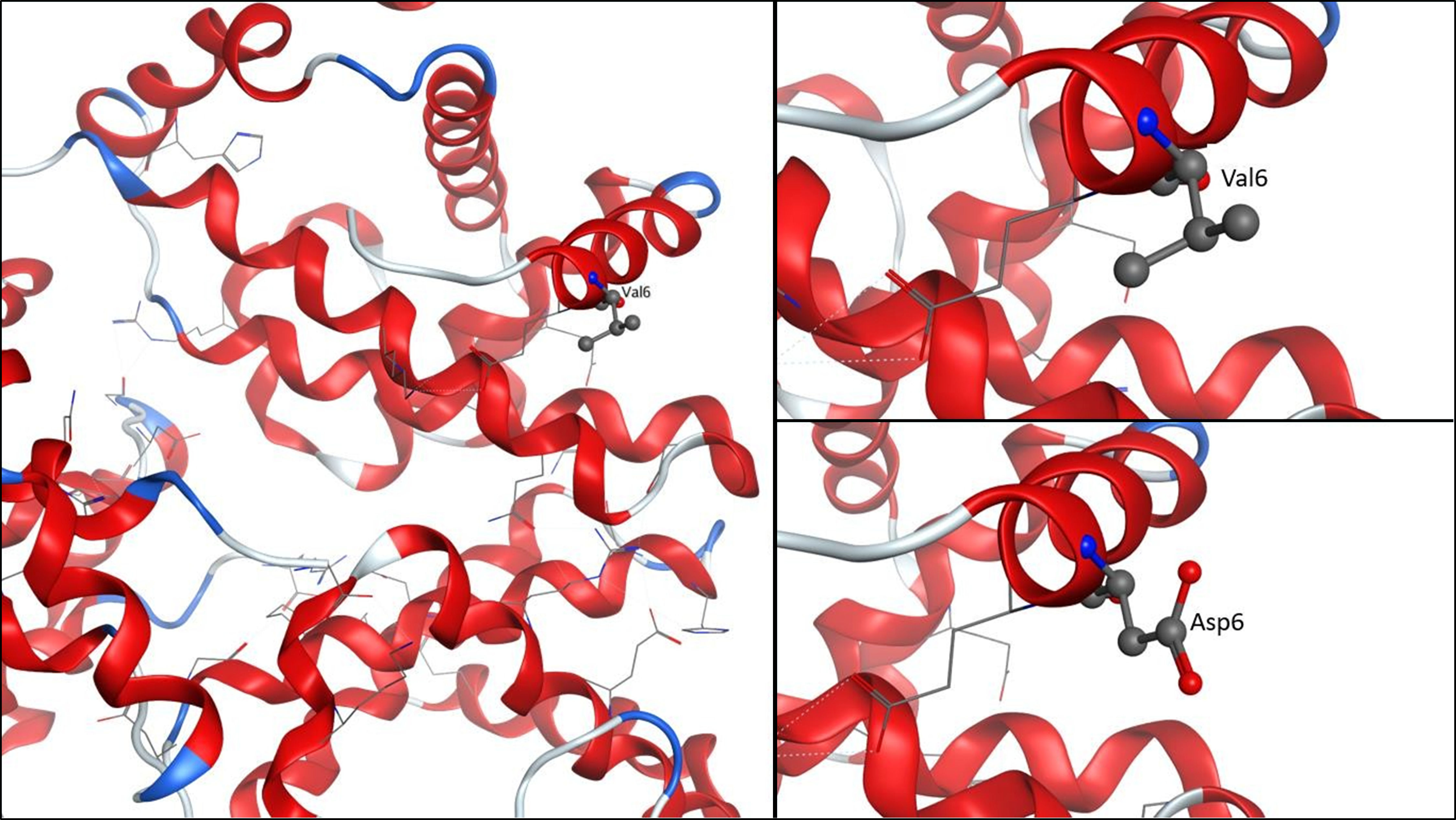 Click for large image | Figure 1. Segment of the β chain of hemoglobin. It shows how the residue at position six is replaced by valine (HbS) and the chain with the aspartic acid residue hemoglobin A (HbA). HbS: hemoglobin S. |
There are additional factors that can positively or negatively influence SCD, including genetic factors such as fetal hemoglobin (HbF) expression and α-thalassemia [29]. It is noteworthy that SCD is theorized to have emerged as a protective mechanism against certain diseases, as it has been reported to confer defense against human immunodeficiency virus-1 infection by activating a complex protein network in response to hemolysis [30]. This protective activity has been attributed to increased iron expression, hypoxia, and interferon-induced innate antiviral factors, as well as the inhibition of overexpressed antiviral genes [31].
Other studies, in contrast, highlight the role of iron overload and ferroptosis in the progression of SCD, affecting vital organs such as the heart, kidneys, skin, brain, lungs, liver, and the blood coagulation system [32, 33]. Certain physiological conditions require excessive amounts of iron, as observed in erythroid hyperplasia within the bone marrow [34]. Iron demand may also increase due to enhanced hemolysis or accelerated turnover of newly formed cells, which translates into decreased hemoglobin levels, hematocrit, and erythrocyte counts [35].
| Oxidative Stress | ▴Top |
Aerobic metabolism generates oxygen-derived free radicals, which are highly reactive compounds capable of inducing oxidative damage to cellular components. This damage plays a fundamental role in the onset and progression of various pathological conditions. Although oxidative stress is conventionally defined as an imbalance between the production of ROS and the organism’s ability to neutralize them through antioxidant defenses, it is important to note that free radicals are not merely undesirable byproducts of metabolism but also fulfill essential physiological roles [36, 37]. During normal biological processes, free radicals participate in critical functions such as cellular signaling and immune responses. However, when their production exceeds the body’s antioxidant capacity, the risk of cellular damage increases, contributing to the development of multiple diseases [38].
Reactive oxygen species (ROS)
The generation of ROS can occur directly or indirectly, through reactions involving oxygen or hydrogen peroxide with iron in the body, such as in Fenton or Haber–Weiss reactions.
Approximately 80% of total body iron is contained within erythrocytes, while the remaining fraction is stored in macrophages, hepatocytes, or within heme groups [38]. Iron plays a key role in various enzymatic and transport processes, where it alternates between the ferric (Fe3+) and ferrous (Fe2+) states. During this redox cycle, iron can donate one of its electrons to a chemical species, resulting in the formation of free radicals. In the cytosol, iron can be present in its reduced form within the so-called labile iron pool [39, 40], which is particularly active in redox reactions and promotes cell death through the generation of reactive species [41, 42].
On the other hand, it is important to highlight the role of macrophages in recycling iron. These cells phagocytose senescent or defective erythrocytes at extravascular sites, such as the spleen, in a process known as erythrophagocytosis [43, 44]. Therefore, macrophages play a fundamental role in iron regulation, as there is no physiological mechanism for the active excretion of iron in humans. Iron absorption and recycling are tightly regulated to maintain iron homeostasis [45]. Among the products of erythrocyte phagocytosis and degradation is iron, which originates from the heme prosthetic group. A transport protein facilitates its translocation from the membrane to the cytosol, where it is catabolized by heme oxygenase (heme oxygenase (HMOX)1/HMOX2), generating biliverdin, CO2, and iron as end products [46, 47]. The released iron is subsequently stored in ferritin and, to a lesser extent, in the form of hemosiderin [48]. When the binding capacity of these proteins is exceeded, they become saturated, which may lead to conditions such as liver damage and iron-induced neurodegeneration due to the presence of free iron.
Iron used for hemoglobin synthesis is exported from macrophages through the transmembrane protein ferroportin-1 [49], which is inhibited by hepcidin, a systemic iron regulator produced by hepatocytes and regulated by intracellular iron levels and oxygen availability [50]. Hepcidin levels, in turn, may be reduced under conditions of chronic inflammation, which require increased red blood cell production through erythropoiesis and, consequently, higher iron demand. The amount of iron required by the body depends on the number of erythroid cells needed at a given time. This iron is primarily supplied by the transport protein transferrin. Additionally, iron can be absorbed by duodenal enterocytes and subsequently bound to plasma transferrin [51], thereby preventing iron from remaining in its free form and generating ROS.
Transferrin transports iron to erythroid cells via transferrin receptor 1 (TfR1) [52] through clathrin-mediated endocytosis. Iron is released within endosomes and transported either to iron storage pools or to the mitochondria via the divalent metal transporter SLC11A2 (solute carrier family eleven member 2) [53]. This entire process can be stimulated by increased erythrocyte destruction due to cellular instability, leading to elevated rates of iron recycling.
Ferroptosis
Iron-mediated ferroptosis is an oxidative form of cell death characterized by the accumulation of lipid peroxides. Ferroptosis has emerged as a contributing factor in several diseases associated with iron overload, including SCD [54]. This cell death pathway is intricately linked to metabolic processes and disruptions in redox homeostasis [55], which may be driven by frequent blood transfusions or increased intestinal iron absorption resulting from reduced hepcidin levels, ultimately leading to secondary iron overload [56]. In ferroptosis, three major research domains converge to define this process: metabolic mechanisms, control of ROS, and iron regulation [57]. Part of this cycle involves intravascular rupture or hemolysis, which causes the release of erythrocyte contents into the bloodstream [55, 58], increasing levels of free iron. This elevation is reflected in urine, where iron concentrations in patients with SCD are markedly higher, particularly during hemolytic crises.
The mechanism of ferroptosis is initiated when excessive hemolysis saturates the binding capacity of hemopexin, the plasma protein responsible for scavenging and removing free heme. As a result, increased heme uptake occurs, followed by excessive catabolism of heme by heme oxygenases, leading to elevated intracellular iron levels (Fig. 2). This iron can participate in Fenton and Haber–Weiss reactions, generating free radicals that react with the allylic hydrogen atoms of polyunsaturated fatty acid chains. This reaction involves hydrogen abstraction from polyunsaturated fatty acids, triggering a chain reaction that ultimately leads to disruption of the erythrocyte plasma membrane and exacerbates hemolysis [55]. Ferroptosis also involves mitochondrial alterations, including mitochondrial contraction, reduction of cristae, and rupture of the outer membrane, resulting in decreased membrane potential and increased permeability. However, the role of mitochondria in ferroptosis is considered secondary [59].
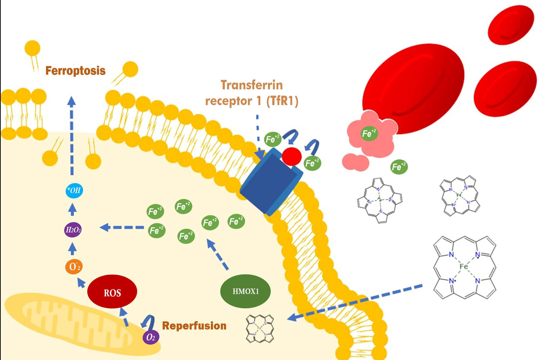 Click for large image | Figure 2. Mechanism of ferroptosis-mediated cell death during hemolysis and ischemia–reperfusion injury in SCD. SCD involves the accumulation of iron-induced lipid peroxides and an increase in ROS, leading to ferroptosis. Excessive hemolysis saturates the capacity of hemopexin, which elevates labile iron through HMOX1 and promotes lipid peroxidation through Fenton reactions. During ischemia–reperfusion, the restoration of blood flow generates an oxidative burst that increases lipid oxidation. In addition, uncoupled nitric oxide synthase, cytochrome P450, and lipoxygenase contribute to the formation of hydroxyl radicals, exacerbating damage to cell membranes. This process culminates in ferroptosis, a form of iron-dependent cell death that compromises membrane integrity and exacerbates tissue damage in SCD. ROS: reactive oxygen species; SCD: sickle cell disease; HMOX1: heme oxygenase 1. |
| HbF | ▴Top |
It has been proposed that HbF may play a role in the regulation of micronutrients and oxidative stress in SCD. A study conducted in Ghana examined the relationship between these factors in children with SCD. The results showed that ambulatory patients had significantly higher HbF levels compared with the control group, whereas hospitalized patients exhibited elevated zinc levels. It has been observed that a persistently high concentration of HbF during postnatal life reduces the tendency for erythrocyte sickling by being excluded from the HbS polymer, thereby improving clinical prognosis due to its higher affinity for oxygen. In addition, HbF can dissociate into dimers that bind to HbS, forming a stable tetramer that does not polymerize.
The study also found a positive correlation between zinc and HbF levels in ambulatory patients. This suggests that both mechanisms, the replacement of aberrant hemoglobin by HbF and increased levels of superoxide dismutase, contribute to the more favorable clinical condition observed in ambulatory patients compared with hospitalized individuals [60]. This effect may reduce hemolysis and, consequently, decrease circulating free iron and ROS, which are key factors in the induction of ferroptosis.
| Hydroxyurea | ▴Top |
The “REACH” trial was a cohort study designed to evaluate the safety, feasibility, and benefits of hydroxyurea administered at the maximum tolerated dose in children from sub-Saharan Africa with SCD and malaria infections. Malaria parasites require iron for the function of iron-sulfur-dependent enzymes, and this mineral is essential for their nucleic acid synthesis. Therefore, body iron stores influence both the rate of infection and the severity of malaria. Hydroxyurea is also known for its antioxidant activity and its ability to chelate iron, playing a crucial role in the prevention of ferroptosis by reducing ROS and lipid peroxidation levels. It has been shown that hydroxyurea not only decreases the incidence of malaria, but also plays an essential role in the management of SCD.
Hydroxyurea induces the production of HbF, which mitigates hemoglobin polymerization and, consequently, reduces vaso-occlusive crises. In addition, its myelosuppressive activity decreases the number of active blood cells, thereby reducing vaso-occlusive and inflammatory events. It is important to note that hydroxyurea also improves nitric oxide bioavailability, promoting vasodilation and reducing ischemic damage in SCD [14]. By chelating iron, hydroxyurea decreases the availability of free iron, a key factor in the induction of ferroptosis. By controlling iron levels and ROS, hydroxyurea helps mitigate oxidative cellular damage, thereby reducing the risk of ferroptosis. Furthermore, its ability to enhance nitric oxide bioavailability promotes vasodilation, which may prevent ischemic injury and reduce the accumulation of lipid peroxides generated during ischemia–reperfusion, a process that exacerbates ferroptosis.
| Hepcidin | ▴Top |
A study conducted in Tanzania aimed to determine whether hepcidin was associated with the severity of anemia in SCD and to investigate factors related to hepcidin and anemia. A total of 199 children from Tanzania, aged between 3 and 18 years, were enrolled. The researchers found that hepcidin levels were lower in children with severe anemia, which was associated with lower steady-state mean hemoglobin levels in the first quartile group. However, this relationship was independent of markers of erythropoiesis or inflammation, although it was associated with an increased reticulocyte percentage [15].
This condition may be linked to elevated levels of erythropoiesis or hypoxia, which override the effect of inflammation and thereby maintain low hepcidin levels. This results in increased iron concentrations, which may activate pathways leading to ferroptosis due to elevated levels of labile free iron. This may also explain why, despite an increased reticulocyte percentage, no increase in mean hemoglobin levels is observed, as these cells may be susceptible to membrane rupture due to oxidative stress. Nevertheless, further studies are required to corroborate these hypotheses.
| Glycosylated Hemoglobin | ▴Top |
Excess iron in the body, associated with SCD, may also introduce bias in other laboratory tests, such as those related to glycemia. Glycated hemoglobin (HbA1c) is widely used for the diagnosis and management of diabetes [16]. However, HbA1c also reflects iron-related erythrocyte properties, which vary by sex. The study showed that both iron and hemoglobin are inversely associated with HbA1c levels but directly associated with glucose levels [17]. Nevertheless, this relationship is more pronounced in women than in men, leading to a tendency to underestimate fasting glucose levels in men compared with women. This may be because men generally have higher hemoglobin and iron levels than women [61].
On the other hand, SCD often increases iron levels due to the need to stimulate erythropoiesis. In addition, excess labile iron may induce red blood cell rupture through ferroptosis, which could reduce HbA1c levels in patients with this disease. For this reason, it is essential to take this parameter into account when performing tests aimed at measuring glycemia in patients with SCD.
| Kidney Damage | ▴Top |
Patients with lower hemoglobin levels (Hb < 7 g/dL) have a higher risk of developing chronic kidney damage. In a study involving more than 16,000 patients with SCD, it was observed that the risk of chronic kidney disease (CKD) decreases significantly as hemoglobin levels increase [62]. This may be due to the high iron demand required for erythrocyte production, which is further intensified by severe anemia and hemolysis, thereby playing a crucial role in the pathogenesis of kidney damage associated with SCD.
In this disease, renal damage primarily arises from persistent hypoxia, oxidative stress, and microvascular injury [63]. The renal medulla, characterized by low oxygen tension and high osmolarity, is particularly susceptible to recurrent vaso-occlusive crises and ischemia–reperfusion injury. These processes trigger endothelial dysfunction, inflammation, and the release of ROS, resulting in detrimental systemic effects [18]. In addition, intravascular hemolysis releases free hemoglobin, intensifying oxidative stress through nitric oxide depletion and increased ROS generation. This toxic environment contributes to multiorgan damage, particularly affecting the renal glomerulus.
A crucial aspect of this damage is iron release and intracellular accumulation. In SCD, hemolysis and erythrophagocytosis release heme iron into the labile iron pool, activating redox reactions that promote lipid peroxidation in glomerular cells. This oxidative damage affects podocytes and mesangial cells, compromising the integrity of the glomerular barrier and favoring the development of albuminuria [64].
Ischemia–reperfusion injury also plays a significant role in the progression of renal damage [65]. During hypoxic episodes, the glomeruli, which are extremely sensitive to these conditions, sustain mitochondrial injury [66]. When blood flow is restored, the rapid reintroduction of oxygen generates a burst of ROS, exacerbating oxidative stress and perpetuating progressive renal dysfunction. Chronic inflammation and the systemic impact of elevated ROS levels and inflammatory cytokines further intensify immune system activation, contributing to renal deterioration. This inflammatory environment, combined with oxidative stress, not only impairs renal function but also aggravates the systemic complications inherent to SCD.
| Ferroportin | ▴Top |
The Q248H mutation of ferroportin (FPN), a key transmembrane protein responsible for iron export and the regulation of iron homeostasis, has a significant impact on processes related to oxidative stress and ferroptosis [67]. This mutation, located in the SLC40A1 gene, confers partial insensitivity to hepcidin, the hormone that regulates ferroportin function. As a result, the mutation leads to continuous iron export, thereby reducing intracellular iron levels in macrophages and other cells involved in iron storage and recycling.
In a study conducted in South Kivu, Democratic Republic of Congo, the Q248H ferroportin mutation was investigated in a pediatric population of 1,071 children under 5 years of age. This mutation, present in approximately 11.4% of the children evaluated, induces partial hepcidin insensitivity of ferroportin, leading to continuous iron export from cells into the plasma and reduced intracellular iron levels [68]. Although this characteristic might appear protective against iron-dependent infections, such as malaria, and against oxidative damage, the results indicated that the mutation did not confer significant protection against anemia or malaria-associated outcomes.
From a ferroptosis perspective, the Q248H ferroportin mutation has complex implications. Under conditions of oxidative stress, such as in SCD, hemolysis and ischemia–reperfusion injury generate elevated levels of labile plasma iron, which increase the production of ROS. These ROS promote lipid peroxidation and ferroptosis-mediated cell death. Although the mutation reduces intracellular iron in certain cells, its net effect under stress conditions may exacerbate dysfunction by facilitating continuous iron transport into extracellular compartments and other susceptible cells.
| Iron Deficiencies | ▴Top |
The population of children under 5 years of age in East Africa represents a particularly vulnerable group due to factors such as poverty, malnutrition, limited access to health services, and the high burden of infectious diseases such as malaria. Anemia data from 2001 to 2021 in countries including Ethiopia, Tanzania, Kenya, and Mozambique reflect significant disparities in the prevalence of mild, moderate, and severe cases. These regional differences can be attributed to factors such as climatic conditions, which influence malaria transmission; health care infrastructure, which limits access to preventive and therapeutic interventions; and food security, which determines the availability of essential micronutrients such as iron and folate [19].
In contrast, the metabolic pathway of ferroptosis is associated with specific biological processes that, while sharing some underlying factors with anemia, operate at a distinct molecular level. In the context of SCD, excessive hemolysis leads to the accumulation of free iron, triggering Fenton reactions that generate ROS and cause lipid peroxidation. This process culminates in ferroptosis, an iron-dependent form of cell death. Unlike nutritional anemia or anemia induced by infections such as malaria, ferroptosis in SCD is driven by iron overload rather than iron deficiency. This highlights that while iron scarcity is the primary cause of anemia in many contexts, in SCD excess iron contributes to significant cellular damage.
| Chronic Hemolysis | ▴Top |
The results of the Realizing Effectiveness Across Continents with Hydroxyurea (REACH) trial highlight the safety, tolerability, and efficacy of hydroxyurea in children with SCA in sub-Saharan Africa [20], while also revealing important links to the metabolic pathway of ferroptosis. Hydroxyurea produced sustained increases in hemoglobin and HbF, while reducing the absolute neutrophil count (ANC), reflecting its ability to partially attenuate the effects of chronic hemolysis and oxidative stress. However, the dose-limiting toxicities (DLTs) reported, such as thrombocytopenia and anemia associated with infections including malaria, underscore the complexity of managing iron overload and ROS generation.
Chronic hemolysis and recurrent infections, exacerbated by malaria, promote the accumulation of labile iron, which catalyzes Fenton reactions that generate ROS and lipid peroxidation hallmark processes of ferroptosis. Although hydroxyurea increases HbF levels, stabilizing erythrocytes and reducing the risk of vaso-occlusion, persistent oxidative stress in high infection burden settings may intensify vulnerability to ferroptosis, particularly in patients with genetic traits such as α-thalassemia.
| Splenomegaly | ▴Top |
The results of the REACH trial, which link splenomegaly and hydroxyurea treatment in children with SCD, can be associated with the metabolic pathway of ferroptosis as an important secondary mechanism. Splenomegaly, associated with the sequestration of damaged erythrocytes and splenic dysfunction, increases the accumulation of free iron derived from hemolysis and erythrophagocytosis, favoring Fenton reactions that generate ROS and lipid peroxidation, both key events in ferroptosis. In addition, hypersplenism observed in these patients, reflected by low hemoglobin and platelet levels, exacerbates systemic oxidative stress, increasing susceptibility to ferroptosis in the spleen and other tissues.
The increased risk of infections such as malaria and the need for frequent transfusions in children with splenomegaly further intensify iron overload, promoting oxidative processes and cellular damage characteristic of ferroptosis. Although hydroxyurea induces a robust HbF response that helps mitigate HbS polymerization, the DLT observed in patients with splenomegaly suggests that oxidative stress and ferroptosis-related effects may influence treatment efficacy and tolerability [21]. These findings underscore the importance of addressing ferroptosis as a secondary therapeutic target through strategies aimed at regulating iron homeostasis and reducing oxidative stress in children with SCD and splenic complications.
| Colombian Context | ▴Top |
A study conducted in Bogota evaluated the frequency of hemoglobin variants according to different regions of the country in a population adjusted for sex and age. This was a retrospective descriptive study of hemoglobin variants analyzed by electrophoresis. The results from 2,224 patients (48.4% men and 51.5% women) showed that 63.3% of patients had no alterations, 14.3% presented thalassemia, 17.3% HbS, 1.8% hemoglobin C (HbC), 0.5% hemoglobin E (HbE), and 0.5% persistent HbF. Regional differences reflected population, environmental, and genetic factors. The Andean region, which accounted for the largest proportion of participants, showed a predominance of normal hemoglobin electrophoresis (63.9%) and a considerable proportion of patients with elevated hemoglobin A2 (HbA2), thalassemia (15.7%), and HbS (15%), the latter in the sickle cell trait (SCT) form hemoglobin AS (HbAS).
In the Caribbean and Pacific regions, a higher prevalence of HbS variants was identified, reaching up to 44.3% in the Pacific region and 26% in the Caribbean region. In the Pacific region, hemoglobinopathies associated with SCA (HbSS, HbS/β-thalassemia) and SCT are predominant, in contrast to the lower proportion of HbS observed in the Andean region (15%). In the Amazon and Orinoquia regions, a marked predominance of normal electrophoresis (> 64%) was observed, with few cases of hemoglobinopathies, particularly thalassemias. Regarding less frequent variants, such as HbC and HbE, these were present at very low proportions across all regions, with HbE being exclusive to the Andean region [22].
Another study conducted in patients with SCD enrolled in the “Ruta de la Vida” program at University Hospital of Valle in Cali, Colombia, reported that most patients originated from Valle del Cauca (75.2%), with significant concentrations in Cali (47.9%) and Buenaventura (16.2%), followed by Nariño (13.1%) and Cauca (11%). The median age at diagnosis was 2.1 years, with 65.4% diagnosed before 3 years of age and 17.0% before 6 months of age. The main clinical conditions reported reflected a high burden of complications related to SCD. Prior to program enrollment, pain was the most common complication (43.8%), followed by hemolytic episodes (32%), acute chest syndrome (13.1%), and splenic sequestration (7.2%). At enrollment, pain (24.2%) and hemolysis (23.5%) remained the most frequent complications, while diagnostic findings included splenomegaly (15.4%), ventricular hypertrophy (3.9%), and avascular necrosis (10.7%) [23].
Additionally, a study examining clinical differences between SCA and SCT in a Colombian population identified patterns of symptoms and progressive complications. Key findings included a high prevalence of pain crises (76.8% overall and 83.7% in SCA), pneumonia, splenomegaly, and cardiac complications. These symptoms were correlated with HbS polymerization, a process that induces erythrocyte deformation, endothelial adhesion, and vascular occlusion. Furthermore, the study showed that 46% of patients with SCA required frequent blood transfusions due to severe hemolysis, whereas only 9.3% of patients with SCT required transfusions, highlighting the disparity in disease severity [23].
The studies analyzed above demonstrate a clear relationship between the pathophysiology of hemoglobinopathies and ferroptosis. The described complications such as vaso-occlusive crises, chronic inflammation, tissue necrosis, and cerebrovascular events reflect the progressive accumulation of cellular damage in tissues exposed to persistent oxidative stress. These studies also highlight that the predominance of HbS variants in specific regions of Colombia, such as the Caribbean and Pacific regions, predisposes affected populations to a higher incidence of complications due to genetic and ancestral factors that amplify inflammatory and cellular damage pathways [22].
Furthermore, ferroptosis may explain the observed associations between pneumonia, splenomegaly, and cardiac complications, in which affected tissues are subjected to systemic inflammation and structural damage. These observations underscore the importance of designing interventions aimed at mitigating ferroptosis, such as the use of targeted antioxidants, lipid peroxidation inhibitors, and iron chelators to limit progressive damage and improve outcomes in patients with hemoglobinopathies. Collectively, these findings reinforce the concept that ferroptosis is not only a central mechanism in the pathophysiology of these diseases but also a key therapeutic target for addressing severe complications and improving the quality of life of affected populations.
| Comparison of SCD in the Colombian and African Contexts | ▴Top |
SCD shows significant differences between Africa and Colombia in terms of prevalence, management, and complications, influenced by genetic, socioeconomic, and health care access factors. In sub-Saharan Africa, where approximately 75% of global cases occur, incidence can reach up to 5% among newborns, particularly in countries such as Ghana, with high infant mortality driven by limited access to medical resources and effective treatments. The most frequent complications include vaso-occlusive crises, severe oxidative stress, and multiorgan damage exacerbated by iron overload and inflammation. Although hydroxyurea has demonstrated efficacy in reducing these complications, its use remains limited due to low availability.
In Colombia, SCD has a higher prevalence in the Caribbean and Pacific regions, where genetic and ancestral factors contribute to rates of up to 44.3% of HbS variants, while the Andean and Amazon regions show lower incidence [22]. Complications include pain crises, severe hemolysis, and acute chest syndrome, with greater severity observed in SCA compared with SCT. Programs such as “Ruta de la Vida” have improved early detection and management; however, limitations in resources and coverage persist. In both settings, greater investment is needed in neonatal screening, access to effective therapies, and strategies to mitigate complications such as oxidative stress and ferroptosis, which severely impact patients’ quality of life.
| Conclusions | ▴Top |
Although ferroptosis is not the primary mechanism in the pathophysiology of SCD, it plays a relevant secondary role that amplifies the clinical complications of this condition. The chronic hemolysis characteristic of SCD releases excess labile iron, triggering Fenton reactions that generate ROS and induce lipid peroxidation. This process exacerbates oxidative damage and contributes to multiorgan complications such as renal injury, chronic inflammation, and vaso-occlusive crises.
In Africa, low hepcidin levels and mutations such as the Q248H ferroportin variant, which facilitates continuous iron export, increase patients’ susceptibility to ferroptosis. In addition, treatments such as hydroxyurea have demonstrated efficacy in reducing ROS, increasing HbF, and chelating free iron, thereby improving clinical outcomes in this population.
In Colombia, clinical cases reflect the predominance of HbS variants in the Caribbean and Pacific regions, which are associated with severe vaso-occlusive crises and renal damage. In these regions, ferroptosis may help explain complications such as chronic inflammation and structural tissue damage in organs exposed to persistent oxidative stress. Studies conducted in Bogotá and Cali highlight the importance of improving early detection and comprehensive management of SCD by addressing not only central mechanisms such as hemolysis, but also secondary factors including ferroptosis.
This analysis underscores the need for therapeutic strategies aimed at regulating iron homeostasis and mitigating oxidative stress in SCD. Interventions such as antioxidants, lipid peroxidation inhibitors, and iron chelators could limit the impact of ferroptosis, significantly improving clinical outcomes and quality of life for patients in both Colombia and Africa.
Acknowledgments
The authors are grateful to Research and Innovation Department of the University Institution Mayor of Cartagena.
Financial Disclosure
This research did not receive any specific grant from funding agencies.
Conflict of Interest
The authors declare that they have no known competing interests.
Author Contributions
Conceptualization: Victor Eljach Cordoba, Sheccid Genes Guerra, Sonia M. Julio Hueto, Jesus D. Rangel Pupo, Jerson A. Zuniga Parra, Neda Alvarez-Ortega, Emilia M. Polo-Carrillo, Jenny Palomares-Bolanos. Data curation: Victor Eljach Cordoba. Formal analysis: Victor Eljach Cordoba, Sheccid Genes Guerra. Funding acquisition: Neda Alvarez-Ortega, Emilia M. Polo-Carrillo, Jenny Palomares-Bolanos. Investigation: Victor Eljach Cordoba, Sheccid Genes Guerra, Sonia M. Julio Hueto, Jesus D. Rangel Pupo, Jerson A. Zuniga Parra, Neda Alvarez-Ortega, Emilia M. Polo-Carrillo, Jenny Palomares-Bolanos. Methodology: Victor Eljach Cordoba, Sheccid Genes Guerra, Sonia M. Julio Hueto, Jesus D. Rangel Pupo, Jerson A. Zuniga Parra, Neda Alvarez-Ortega. Project administration: Neda Alvarez-Ortega, Emilia M. Polo-Carrillo, Jenny Palomares-Bolanos. Resources: Neda Alvarez-Ortega, Emilia M. Polo-Carrillo, Jenny Palomares-Bolanos. Supervision: Neda Alvarez-Ortega, Emilia M. Polo-Carrillo, Jenny Palomares-Bolanos. Validation: Neda Alvarez-Ortega, Emilia M. Polo-Carrillo, Jenny Palomares-Bolanos. Writing – original draft: Victor Eljach Cordoba, Sheccid Genes Guerra, Sonia M. Julio Hueto, Jesus D. Rangel Pupo, Jerson A. Zuniga Parra, Neda Alvarez-Ortega, Emilia M. Polo-Carrillo, Jenny Palomares-Bolanos. Writing – review and editing: Neda Alvarez-Ortega, Emilia M. Polo-Carrillo, Jenny Palomares-Bolanos.
Data Availability
The authors declare that data supporting the findings of this study are available within the article.
| References | ▴Top |
- Saleki K, Rezaei N. Introduction to hematological disorders. Comprehensive Hematology and Stem Cell Research. Elsevier. 2024;1:1-50.
- Johnson CS. The sickle-cell diseases. Reference Module in Biomedical Sciences: Elsevier. 2024.
doi - Pham PTT, Pham PCT, Lew SQ. Chapter 50 sickle cell disease. Renal Disease. Academic Press. 2020;2:813-830.
doi - Inusa BPD, Hsu LL, Kohli N, Patel A, Ominu-Evbota K, Anie KA, Atoyebi W. Sickle cell disease-genetics, pathophysiology, clinical presentation and treatment. Int J Neonatal Screen. 2019;5(2):20.
doi pubmed - Wonkam A, Munung NS, Guindo A, Nnodu O. Priorities for sickle cell disease global research and implementation. Lancet. 2024;404(10465):1790-1792.
doi pubmed - Obadina M, Morris S, Alin T, LeVarge B, Little JA. High-altitude hypoxia is common in adults with sickle cell disease. Blood. 2023;142:3894.
doi - Colombatti R, Birkegard C, Medici M. PB2215: global epidemiology of sickle cell disease: a systematic literature review. Hemasphere. 2022;6:2085-2086.
doi - Collaborators GBDSCD. Global, regional, and national prevalence and mortality burden of sickle cell disease, 2000-2021: a systematic analysis from the Global Burden of Disease Study 2021. Lancet Haematol. 2023;10(8):e585-e599.
doi pubmed - Kumar R, Shanmugam R, Das A. Sickle cell disease in India: prevention-driven social and public health implications. Clinical Epidemiology and Global Health 15. 2022.
- Esoh K, Wonkam-Tingang E, Wonkam A. Sickle cell disease in sub-Saharan Africa: transferable strategies for prevention and care. Lancet Haematol. 2021;8(10):e744-e755.
doi pubmed - Asare EV, Wilson I, Benneh-Akwasi Kuma AA, Dei-Adomakoh Y, Sey F, Olayemi E. Burden of sickle cell disease in Ghana: the Korle-Bu experience. Adv Hematol. 2018;2018:6161270.
doi pubmed - Belvis D, Henderson KJ, Benzon HA. Chapter 38-sickle cell disease. Essentials of Pain Medicine. Elsevier. 2019;4:333-801.
- Nnodu OE, Oron AP, Sopekan A, Akaba GO, Piel FB, Chao DL. Child mortality from sickle cell disease in Nigeria: a model-estimated, population-level analysis of data from the 2018 Demographic and Health Survey. Lancet Haematol. 2021;8(10):e723-e731.
doi pubmed - Olupot-Olupot P, Tomlinson G, Williams TN, Tshilolo L, Santos B, Smart LR, McElhinney K, et al. Hydroxyurea treatment is associated with lower malaria incidence in children with sickle cell anemia in sub-Saharan Africa. Blood. 2023;141(12):1402-1410.
doi pubmed - Lee N, Makani J, Tluway F, Makubi A, Armitage AE, Pasricha SR, Drakesmith H, et al. Decreased hepcidin levels are associated with low steady-state hemoglobin in children with sickle cell disease in Tanzania. EBioMedicine. 2018;34:158-164.
doi pubmed - Ximbi S, Chale-Matsau B, Pillay TSP. Seasonal variation of glycated haemoglobin (HbA1c) in temperate South Africa. Clinica Chemical Acta. 2024;558:119792.
- Kweka B, Lyimo E, Kidola J, Filteau S, Friis H, Manjurano A, et al. Validity of HbA1c in diagnosing diabetes among people with sickle cell trait in Tanzania. Blood. 2019;134:4852.
- Amarapurkar P, Roberts L, Navarrete J, El Rassi F. Sickle cell disease and kidney. Adv Chronic Kidney Dis. 2022;29(2):141-148.e141.
doi pubmed - Hailu BA. Mapping, trends, and factors associated with anemia among children aged under 5 y in East Africa. Nutrition. 2023;116:112202.
doi pubmed - Aygun B, Tomlinson GA, McGann PT, Tshilolo L, Williams TN, Olupot-Olupot P, et al. Optimizing hydroxyurea therapy with reduced laboratory monitoring for children with sickle cell anemia in Sub-Saharan Africa: the reach experience. Blood. 2020;136:17.
doi - Tshilolo L, Tomlinson GA, McGann PT, Latham TS, Olupot-Olupot P, Santos B, et al. Splenomegaly in children with sickle cell anemia receiving hydroxyurea in sub-Saharan Africa. Blood. 2019;134:993.
doi - Vargas-Hernandez DA, Uscategui-Ruiz AC, De Avila J, Romero-Sanchez C. Differences in the distribution of hemoglobin variants according to the geographic regions in a Colombian population. Hematol Transfus Cell Ther. 2023;45(Suppl 2):S140-S147.
doi pubmed - Soto SM, Portilla CA, García-Perdomo HA. Clinical and demographic characteristics of sickle cell disease in Colombian children. Hematology in Clinical Practice. 2023;14:46-51.
doi - Hu C, Shen L, Zou F, Wu Y, Wang B, Wang A, Wu C, et al. Predicting and overcoming resistance to CDK9 inhibitors for cancer therapy. Acta Pharm Sin B. 2023;13(9):3694-3707.
doi pubmed - Pollock G, Negre O, Ribeil JA. Gene-addition/editing therapy in sickle cell disease. Presse Med. 2023;52(4):104214.
doi pubmed - Camacho RA, Machado AV, de Oliveira Mendonca F, Teixeira-Alves LR, Guimaraes-Nobre CC, Mendonca-Reis E, da Silva PF, et al. Unraveling DEHP influence on hemoglobin S polymerization in sickle cell disease: Ex vivo, in vitro and in silico analysis. Toxicol In Vitro. 2024;98:105832.
doi pubmed - Henry ER, Cellmer T, Dunkelberger EB, Metaferia B, Hofrichter J, Li Q, Ostrowski D, et al. Allosteric control of hemoglobin S fiber formation by oxygen and its relation to the pathophysiology of sickle cell disease. Proc Natl Acad Sci U S A. 2020;117(26):15018-15027.
doi pubmed - Raghuraman A, Lawrence R, Shetty R, Avanthika C, Jhaveri S, Pichardo BV, Mujakari A. Role of gene therapy in sickle cell disease. Dis Mon. 2024;70(7):101689.
doi pubmed - Khandros E, Blobel GA. Elevating fetal hemoglobin: recently discovered regulators and mechanisms. Blood. 2024;144(8):845-852.
doi pubmed - Kumari N, Nouraie M, Ahmad A, Lassiter H, Khan J, Diaz S, Afangbedji N, et al. Restriction of HIV-1 infection in sickle cell trait. Blood Adv. 2021;5(23):4922-4934.
doi pubmed - Kumari N, Ahmad A, Berto-Junior C, Ivanov A, Wen F, Lin X, Diaz S, et al. Antiviral response and HIV-1 inhibition in sickle cell disease. iScience. 2024;27(2):108813.
doi pubmed - Li H, Kazmi JS, Lee S, Zhang D, Gao X, Maryanovich M, Torres L, et al. Dietary iron restriction protects against vaso-occlusion and organ damage in murine sickle cell disease. Blood. 2023;141(2):194-199.
doi pubmed - Nasimuzzaman M, Malik P. Role of the coagulation system in the pathogenesis of sickle cell disease. Blood Adv. 2019;3(20):3170-3180.
doi pubmed - Lins CF, Salmon CEG, Amorim de Souza L, Quesado RCS, de Souza Moraes R, Silva-Pinto AC, Matos MA, et al. Quantitative MRI evaluation of bone marrow in sickle cell disease: relationship with haemolysis and clinical severity. Clin Radiol. 2023;78(3):e268-e278.
doi pubmed - Vallelian F, Buehler PW, Schaer DJ. Hemolysis, free hemoglobin toxicity, and scavenger protein therapeutics. Blood. 2022;140(17):1837-1844.
doi pubmed - Bajaj S, Singh S, Sharma P. Chapter 19: role of antioxidants. Nutraceutical fruits and foods for neurodegenerative disorders. Academic Press. 2024;353-378.
- Oztas Y, Bosgelmez II. Chapter 6: Oxidative stress. Sickle cell disease and emerging roles for antioxidants in treatment strategies. Pathology. 2020;65-75.
- Dutt S, Hamza I, Bartnikas TB. Molecular mechanisms of iron and heme metabolism. Annu Rev Nutr. 2022;42:311-335.
doi pubmed - Nolfi-Donegan D, Pradhan-Sundd T, Pritchard KA, Jr., Hillery CA. Redox signaling in sickle cell disease. Curr Opin Physiol. 2019;9:26-33.
doi pubmed - Shakya A, McKee NW, Dodson M, Chapman E, Zhang DD. Anti-Ferroptotic effects of Nrf2: beyond the antioxidant response. Mol Cells. 2023;46(3):165-175.
doi pubmed - Roemhild K, von Maltzahn F, Weiskirchen R, Knuchel R, von Stillfried S, Lammers T. Iron metabolism: pathophysiology and pharmacology. Trends Pharmacol Sci. 2021;42(8):640-656.
doi pubmed - Alayash AI. Oxidative pathways in the sickle cell and beyond. Blood Cells Mol Dis. 2018;70:78-86.
doi pubmed - Borges MD, Sesti-Costa R. Macrophages: key players in erythrocyte turnover. Hematol Transfus Cell Ther. 2022;44(4):574-581.
doi pubmed - Li G, Qiang Y, Li H, Li X, Dao M, Karniadakis GE. In silico and in vitro study of the adhesion dynamics of erythrophagocytosis in sickle cell disease. Biophys J. 2023;122(12):2590-2604.
doi pubmed - Ginzburg YZ, Vinchi F. Chapter 72: iron overload. Transfusion medicine and hemostasis. Elsevier. 2019;3:433-436.
- Wang X, Zhang S, Xue D, Neculai D, Zhang J. Metabolic reprogramming of macrophages in cancer therapy. Trends Endocrinol Metab. 2025;36(7):660-676.
doi pubmed - Humar R, Schaer DJ, Vallelian F. Erythrophagocytes in hemolytic anemia, wound healing, and cancer. Trends Mol Med. 2022;28(11):906-915.
doi pubmed - Jayakumar D, KK SN, Periandavan K. Triad role of hepcidin, ferroportin, and Nrf2 in cardiac iron metabolism: From health to disease. J Trace Elem Med Biol. 2022;69:126882.
doi pubmed - Camaschella C, Nai A, Silvestri L. Iron metabolism and iron disorders revisited in the hepcidin era. Haematologica. 2020;105(2):260-272.
doi pubmed - Elbostany EA, Elghoroury EA, Thabet EH, Rashad AA, Rasheed EA, El-Saeed GSM, Abdelhalim DA, et al. Oxidative stress and hepcidin expression in pediatric sickle cell anemia with iron overload. Hematol Oncol Stem Cell Ther. 2023;16(3):238-244.
doi pubmed - Anderson GJ, Lu Y, Frazer DM, Collins JF. Iron; Intestinal absorption. Encyclopedia of Gastroenterology. Oxford. 2020;2:301-311.
- Donaldson JG, Schaffer JE. Transport and secretion, endocytosis. encyclopedia of biological chemistry III. Oxford. 2021;3:451-454.
- Andrews NC. Chapter 78: iron absorption. Physiology of the Gastrointestinal Tract. Burlington. 2006;1983-1992.
- Fortuna V, Lima J, Oliveira GF, Oliveira YS, Getachew B, Nekhai S, Aschner M, et al. Ferroptosis as an emerging target in sickle cell disease. Curr Res Toxicol. 2024;7:100181.
doi pubmed - Fan CH, Zeng XQ, Feng RM, Yi HW, Xia R. Comprehensive review of perioperative factors influencing ferroptosis. Biomed Pharmacother. 2024;179:117375.
doi pubmed - Liu G, Xie X, Liao W, Chen S, Zhong R, Qin J, He P, et al. Ferroptosis in cardiovascular disease. Biomed Pharmacother. 2024;170:116057.
doi pubmed - Stockwell BR. Ferroptosis turns 10: Emerging mechanisms, physiological functions, and therapeutic applications. Cell. 2022;185(14):2401-2421.
doi pubmed - Arnhold J. Chapter 5: Mechanisms of cell death. Cell and Tissue Destruction. 2020;135-53.
- Wang H, Liu C, Zhao Y, Gao G. Mitochondria regulation in ferroptosis. Eur J Cell Biol. 2020;99(1):151058.
doi pubmed - Owusu-Poku AG, Gyamfi D, Togbe E, Opoku S, Ephraim RKD, Asibey JG, et al. Interplay between fetal hemoglobin, micronutrients and oxidative stress biomarkers in sickle cell anemia children. Human Nutrition & Metabolism. 2022;30:200173.
doi - Alzahrani BA, Salamatullah HK, Alsharm FS, Baljoon JM, Abukhodair AO, Ahmed ME, Malaikah H, et al. The effect of different types of anemia on HbA1c levels in non-diabetics. BMC Endocr Disord. 2023;23(1):24.
doi pubmed - Ershler WB, De Castro LM, Pakbaz Z, Moynahan A, Weycker D, Delea TE, Agodoa I, et al. Hemoglobin and end-organ damage in individuals with sickle cell disease. Curr Ther Res Clin Exp. 2023;98:100696.
doi pubmed - Inusa BPD, Liguoro I, Tayo B, Booth C, Turner C, Dalton NR. Reliability of different estimated glomerular filtration rate as measures of renal function in children with sickle cell disease. Blood Cells Mol Dis. 2021;91:102590.
doi pubmed - Derebail VK, Ciccone EJ, Zhou Q, Kilgore RR, Cai J, Ataga KI. Progressive decline in estimated GFR in patients with sickle cell disease: an observational cohort study. Am J Kidney Dis. 2019;74(1):47-55.
doi pubmed - Castro-Sesquen YE, Saraf SL, Gordeuk VR, Nekhai S, Jerebtsova M. Use of multiple urinary biomarkers for the early detection of chronic kidney disease in sickle cell anemia. Blood Adv. 2023;7(11):2606-2608.
doi pubmed - Liem RI, Lanzkron S, T DC, DeCastro L, Desai AA, Ataga KI, Cohen RT, et al. American Society of Hematology 2019 guidelines for sickle cell disease: cardiopulmonary and kidney disease. Blood Adv. 2019;3(23):3867-3897.
doi pubmed - Saraf SL, Gordeuk VR. Chapter 5 - Iron. Essential and Toxic Trace Elements and Vitamins in Human Health. 2020;83-102.
- Bahati YL, Delanghe J, Balaluka GB, Vandepoele K, Cirhuza JC, Kishabongo AS, Philippe J. Ferroportin Q248H mutation was not found to be protective against malaria and anemia in children under 5 years living in South Kivu/Democratic Republic of Congo, an endemic area of P lasmodium infection. Heliyon. 2022;8(9):e10460.
doi pubmed
This article is distributed under the terms of the Creative Commons Attribution 4.0 International License (CC BY 4.0), which permits unrestricted use, distribution, and reproduction in any medium, including commercial use, provided the original work is properly cited.
Journal of Hematology is published by Elmer Press Inc.